
Page 95 - 2019_09-HaematologicaMondo-web
P. 95
Thiazolidinones reduce iron overload
bone marrow cells from Wt Balb/C mice after the admin- istration of the compounds (P<0.05) (Figure 4A). These results suggest that the investigated compounds might also target Erfe, Gdf15 and Twsg1 in bone marrow ery- throid cells to promote hepatic hepcidin expression, and also imply that erythropoietin may not be the only regu- lator of Erfe, Gdf15 and Twsg1. Subsequently, we analyzed the levels of Gdf15, Twsg1 and Erfe mRNA expression in bone marrow cells from Hbbth3/+ mice through qRT-PCR. As shown in Figure 4B, Gdf15, Twsg1 and Erfe mRNA lev-
AB
els were significantly diminished (P<0.05), in line with the results observed in Wt Balb/C mice (Figure 4A). These results demonstrated that the agonists also targeted ery- throid regulators to elevate hepcidin expression, even though the precise molecular mechanisms warrant further detailed investigation.
Hepcidin deficient (Hamp1-/-) mice were resistant to iron changes induced by compounds 93, 156 and 165
To verify that hepatic hepcidin is the iron-relevant target
CD
E
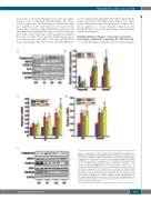
Figure 3. Compounds 93, 156 and 165 targeted SMAD1/5/8 signaling. (A) P- SMAD1/5/8, P-ERK1/2 and TMPRSS6 levels determined by western blot analy- sis of liver specimens from 8-week old Balb/C mice 24 h after administration of compounds 93, 156 and 165 at a dose of 30 mg/kg body weight. (B) Changes of TMPRSS6 and downstream target genes of P-SMAD1/5/8 signaling: Id1 and Smad7 determined by quantitative reverse transcriptase polymerase chain reaction analysis (qRT-PCR) (n=4-6) in liver specimens of these mice. (C) Changes of hepcidin mRNA in Hepa 1-6 cells at the indicated times after treat- ment with compounds 93, 156 and 165 at a concentration of 10 μM (n=4-6). (D) Changes of Id1 and Samd7 were determined by qRT-PCR analysis (n=4) of Hepa 1-6 cells 24 h after treatment with compounds 93, 156 and 165 (10 μM). (E) Variations of P-SAMD1/5/8, SMAD1, P-ERK1/2, ERK1/2 and TMPRSS6 lev- els analyzed by western blot in Hepa 1-6 cells 24 h after treatment with com- pounds 93, 156 and 165 at a concentration of 10 μM. *P<0.05; #P<0.001, rel- ative to untreated control (Ctrl).
haematologica | 2019; 104(9)
1773
