
Page 22 - 2019_09-HaematologicaMondo-web
P. 22
Editorials
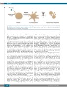
Figure 1. An illustration of thrombin-induced effects on platelets over time. Thrombin produced through coagulation binds to and activates platelet receptors, lead- ing to platelet activation, degranulation, and filopodia formation. At a certain time after the hemostatic plug has contracted, platelets break down into membrane- bound fragments of defined sizes through a distinct mechanism.
1700
happens to platelets after activation, and how they may play a role in hemostasis or clearance. This paradigm shift may help to elucidate novel mechanisms of platelet behav- ior and clarify the functional roles of such fragments of pre- viously activated platelets.
Using transmission and scanning electron microscopy, Kim et al. observed that after 15 min of thrombin treatment, platelets broke up into separate membrane-bound particu- lates that contained granules, mitochondria, and vacuoles. Remarkably, this fragmentation was not seen after expo- sure to other platelet agonists such as collagen or ADP, although the platelets exhibited morphological changes associated with activation such as filopodia formation and spreading. This finding suggests that platelet activation cre- ates agonist-specific behavior, and only when thrombin is being generated can platelets start to break up into frag- ments.
As actin is necessary for cytoskeletal rearrangement and filopodia extension,9 the authors investigated localization of actin after exposure to thrombin. After fragmentation, actin was retained in the particulates, and gradually disappeared as the fragments became smaller. It is also worthy of note that intracellular levels of calcium correlated with fragmen- tation, such that calcium levels dropped as the platelets dis- integrated. Mitochondrial function also decreased as the platelets fragmented. Mitochondria appeared to translocate to the periphery of the cell, or even escape to the extracel- lular space. Clot contraction force plateaued and the gener- ation of reactive oxygen species coincided with the initia- tion of platelet fragmentation, suggesting that clot contrac- tion is stopped by the loss of cellular energy in the form of ATP due to mitochondrial dysfunction. Actin-myosin is an ATP-driven motor, and these results support the previously seen loss of the actin-regulated cytoskeleton.
The authors noted that platelet fragmentation is different from typical necrosis, as fragmentation seemed to be a reg- ulated process that did not entail cellular rupture as the membranes remained intact. To assess whether these platelet fragments were due to apoptotic signaling, caspase activity was assessed. Surprisingly, platelets exposed to up
to 5 U/mL thrombin did not appear to activate caspases. If this is not apoptosis, the question is what is responsible for platelet fragmentation? The authors identified calpain, a cysteine protease that is believed to recognize tertiary struc- ture as a cleavage site instead of sequence-specific activity,10 as one enzyme responsible for these processes. Interestingly, maximal calpain activity coincided with the initiation of fragmentation and functional mitochondrial loss. ALLN, a calpain inhibitor, was able to delay thrombin- induced fragmentation and inhibit mitochondrial loss. However, ALLN was unable to inhibit calpain cleavage products completely. Also, it is clear that inhibition of cal- pains alone is not enough to prevent platelet fragmentation but the observations suggest that proteases are vital for the fragmentation process to occur.
While Kim et al. provided an elegant in vitro characteriza- tion and outlined the mechanism of platelet fragmentation, the biological significance of this process awaits further elu- cidation. For instance, do these platelet fragments play a role in the breakdown of platelet-rich clots, and can aber- rant fragmentation play a role in thrombosis? Moreover, it is worth noting the time delay in fragmentation upon treat- ment with thrombin and after platelet contraction, which may be significant to its function. While the GPIb-IX-V receptor complex does not typically lead to a fast and strong intracellular signal, protease-activated receptors, like most G protein-coupled receptors, can rapidly induce full activa- tion of platelets. Thus, what is the mechanism in the platelet that causes fragmentation to proceed only after platelet activation and contraction events have run their courses? The authors demonstrated that force and time are likely important factors in the process of fragmentation (Figure 1). It would be extremely interesting to understand how signaling and cytoskeletal proteins in the platelet respond to these forces and temporal factors. Furthermore, perhaps there is a balance between traditional apoptotic pathways and fragmentation during exposure to a combi- nation of agonists. When would a platelet undergo apopto- sis rather than fragmentation in response to multiple ago- nists? Finally, it remains to be addressed how platelet frag-
haematologica | 2019; 104(9)
