
Page 77 - 2019_08-Haematologica-web
P. 77
Guadecitabine in selected MDS and AML after azacitidine failure
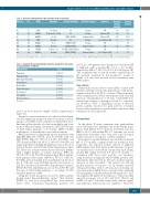
Table 2. Baseline characteristics and outcome of the responders.
Patient n. Sex
#2F
#4 M
#9 F
#10 M
#21 M #24 M #29 M
#53 M
WHO
AML
RAEB-1 RAEB-1 AML RAEB-2 RAEB-2 RCMD
RAEB-2
Karyotype
+8, del20q
Del1p,del5q, del11q +1, +21 Normal
-Y, +8 Normal Del20q, +21
Normal
Somatic mutations
No
No
PHF6, RUNX1
No
SETBP1, RIT1
BCOR, STAG2
RUNX1, U2AF1, ASXL1
EZH2, SETBP1
AZA first response
Relapse
Relapse
Primary failure
Relapse
Relapse
Primary failure
Relapse
Primary failure
Response Response duration (months)
Survival (months)
CR 13* 42**
Marrow CR 19* 19
Marrow CR 21* 24** HI + Marrow CR 10 10 PR 9 14
CR 31* 34**
HI 7 10 HI + Marrow CR 8 12
*Relatively long-term responders. **Relatively long-term survivors. F: female; M: male; WHO: World Health Organization; AZA: azacitidine; RCMD: refractory cytopenia with mul- tilineage dysplasia; CMML: chronic myelomonocytic leukemia; RAEB: refractory anemia with excess blasts; RAEB-t: RAEB in transformation; AML: acute myeloid leukemia.
Table 3. Grade III-IV non-hematologic toxicities during first nine cycles of guadecitabine treatment.
x109/L, 21 of 49 patients were classified as low-risk and 28 as high-risk, with a median OS of 9.2 vs. 5.7 months, respectively (HR=1.7, 95%CI: 0.8-3.8; P=0.16) (Figure 4B). This compared with 11 and 4.5 months, respectively, for the patients included in the prognostic model of Nazha et al. who had received various treatments after HMA failure.
Side effects
Ninety-nine serious adverse events (SAE) occurred in 44 patients, and they were mostly hematologic, with myelo- suppression in 88 of 99 (88%) of events. Thirteen patients were hospitalized for febrile neutropenia with a median duration of hospitalization of 14 days. Grade III-IV non- hematologic toxicities occurring in at least 3% of patients are shown in Table 3. Regarding toxicity at injection points, patients reported less pain and less secondary lesions with subcutaneous guadecitabine injections than with previous AZA injections.
Discussion
In this phase II study, treatment with guadecitabine after AZA failure was generally safe in this elderly popu- lation, with limited dose reductions. It yielded a modest ORR of 14.3% and median OS of 7.1 months, but a few longer-term responders were seen, and some biological prognostic factors of response could be identified.
Responders to guadecitabine in our study had a median OS of 17.9 months, compared with six months in non- responders, the reported median survival of high-risk MDS patients after AZA failure in the literature.2 In previ- ous smaller series of decitabine salvage after AZA failure, median OS ranged between 5.9 and 11.8 months.2,15,16 Compared with those series, and also our experience using decitabine in high-risk MDS/CMML patients after AZA failure (that reported no CR and a median response duration of only 3 months),17 the current two CR and median duration of response of 11.5 months achieved with guadecitabine, with 4 of 8 responses exceeding one year, may appear slightly better. A recent study comparing a 5-day regimen of guadecitabine (60-90 mg/m2/d) to a 10- day regimen (60 mg/m2/d) in relapsed or refractory AML reported a response rate of 16% and 30.2% (P=0.1), and an OS of 5 and 7.1 months, respectively, with no signifi- cant difference between the two regimens.18 However,
Patients N=23
Pulmonary
Cardiovascular Musculary/denutrition Transaminase
Renal failure Gastro-intestinal Neurological Uro-genital Endocrinological
N: number.
N (%)
7 (12.5%)
4 (7.1%) 4 (7.1%) 4 (7.1%) 3 (5.3%) 3 (5.3%) 2 (3.6%) 2 (3.6%) 2 (3.6%)
cycle 1 in bone marrow samples (Online Supplementary Figure S3).
Except for somatic mutations, no other baseline param- eter had significant prognostic value for response, includ- ing age, sex, ECOG status, transfusion dependency, base- line hemoglobin, platelet, absolute neutrophil count, bone marrow blast percentage, cytogenetics, IPSS, IPSS-R, type of AZA failure (primary or secondary), LINE1 baseline methylation, or demethylation rate with treatment.
Overall survival was significantly shorter in patients with high IPSS (HR=1.81, 95%CI: 1.1-2.97; P=0.02), with very high IPSS-R (HR=1.5, 95%CI: 1.2-1.87; P=0.0004), and TP53 mutation (HR=2.23, 95%CI: 1.09-4.57; P=0.028), and longer in patients with high demethylation rate in blood on day 8 of the first cycle (P=0.02) (Online Supplementary Figure S4). There was a trend towards shorter survival in patients with a higher number of somatic mutations (HR=1.18, 95%CI: 0.97-1.44; P=0.099) and prolonged OS in patients with primary AZA failure (HR=0.51, 95%CI: 0.25-1.01; P=0.054), and low baseline level of methylation in blood on day 8 of the first cycle (P=0.066) and in bone marrow on day 28 of the first cycle (P=0.083). In multivariate analysis, IPSS-R (P=0.03), demethylation rate in blood (P=0.03) and the type of AZA failure (primary vs. secondary; P=0.01) remained predictive of OS.
Using the recent prognostic model for MDS patients having failed hypomethylating agents15 that includes ECOG >1, very poor cytogenetics, age, bone marrow blasts >20%, transfusion dependency, platelets <30
haematologica | 2019; 104(8)
1569
