
Page 41 - 2019_08-Haematologica-web
P. 41
MRD in AML
mortality.4 Although improvements in genomic classifica- tion of AML have refined our current risk stratification systems, relapses are unfortunately still common even with risk-adapted treatment.
Assessment of measurable residual disease (MRD), also called “minimal residual disease,” allows for the detection and quantification of lower levels of residual leukemia that can be detected by morphological assessment alone.5,6 In both pediatric and adult acute lymphoblastic leukemia, MRD is routinely used to refine prognostic assessment and also allocate post-remission therapies, particularly in the pediatric population.7-11 Similarly, studies utilizing var- ious methods of MRD determination have consistently shown that MRD is highly prognostic in AML and com- plements (and sometimes supersedes) historically relevant pretreatment characteristics.12-18 However, many questions remain as to the optimal MRD assay, appropriate timing of MRD assessment, and whether MRD should guide treatment. Herein, we review methods of MRD detection and the prognostic impact of MRD across AML subtypes. We also offer some practical considerations for MRD test- ing in the clinical setting and review the available data on how MRD can potentially guide post-remission treatment decisions in AML.
Methods of measurable residual disease assessment
Several methods of MRD assessment are available in the clinical and research settings, each of which has its own advantages and disadvantages. The European
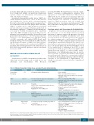
LeukemiaNet MRD Working Party has released compre- hensive, consensus recommendations regarding their appropriate use and technical limitations.6 These various MRD methods vary in their level of sensitivity, applicabil- ity to the vast majority of patients with AML versus only certain subtypes, cost, and level of technical expertise needed to obtain an accurate result, all of which may influence the choice of assay for a particular patient.19 The major differences among MRD technologies are summa- rized in Table 1.
Karyotype analysis and fluorescence in situ hybridization In patients with an abnormal pretreatment karyotype, the persistence of an abnormal leukemia-associated karyotype at remission suggests the presence of residual disease. Persistent cytogenetic abnormalities have been associated with worse survival in several studies and may also identify patients who could benefit from HSCT in first remission.20,21 While cytogenetic analysis at remission adds prognostic information in patients who achieve com- plete remission, the sensitivity of this method is relatively poor, as it can only detect one abnormal metaphase out of 20 (i.e. sensitivity ≈5%). Furthermore, up to 50% of adult patients present with cytogenetically normal AML, further limiting karyotype analysis as a universal MRD marker.1 Fluorescence in situ hybridization may also be used to detect persistent cytogenetic abnormalities with slightly increased sensitivity compared to conventional cytogenet- ics. However, the maximum sensitivity achieved with flu- orescence in situ hybridization is approximately 1%, which is not adequate to detect low levels of clinically rel-
Table 1. Methods of measurable residual disease assessment in acute myeloid leukemia.
evant MRD.22
Method
Conventional karyotyping
FISH
MFC for LAIP or DfN
RT-qPCR
NGS
Sensitivity
~5%
Up to 10-2
10-3 to 10-5
10-4 to 10-6
Highly variable (1% to 10-6)
Advantages
• Common in routine clinical practice
• Useful for numeric cytogenetic abnormalities (i.e. gains or deletions)
• Sensitive
• Fast (results usually available within 24 hours) • Relatively inexpensive
• Applicable to >90% of AML cases
• Sensitive
• Well standardized
• Can be run in any laboratory with RT-qPCR
capabilities
• Potential for very high sensitivity (depending on technology)
• Can test multiple genes at once
Disadvantages
• Poor sensitivity
• Time-consuming and labor-intensive
• Applicable only to patients with baseline abnormal
karyotype (~50%)
• Worse sensitivity than MFC or PCR
• Applicable only to patients with baseline abnormal
karyotype (~50%)
• Potential for immunophenotypic shifts (mitigated by using a DfN-based approach)
• Requires significant technical expertise to interpret
• Limited standardization across laboratories
• Appropriate molecular targets present in <50% of cases (<35% in older adults)
• Many mutations are not suitable for MRD detection (e.g. FLT3)
• Time-consuming and labor-intensive
• Results may take several days
• Low sensitivity with most commonly used platforms • May be confounded by persistence of preleukemic
mutations (e.g. CHIP)
• Results may take several days
• Expensive
• Not standardized
• Requires complex bioinformatics
FISH: fluorescence in situ hybridization; MFC: multiparameter flow cytometry; PCR: polymerase chain reaction; LAIP: leukemia-associated immunophenotypes; DfN: difference from normal; AML: acute myelod leukemia; RT-qPCR: real-time quantitative polymerase chain reaction; NGS: next-generation sequencing; MRD: measurable residual disease; CHIP: clonal hematopoiesis of indeterminate potential.
haematologica | 2019; 104(8)
1533
