
Page 79 - 2019_07 resto del Mondo-web
P. 79
Post-lenalidomide immunomodulation in del(5q) MDS
Illumina next-generation sequencing and data analysis
NGS and demultiplexing were performed on an Illumina MiSeq sequencer (600-cycle single indexed, paired-end run, V3- chemistry). Analysis of rearranged IGH and TRB loci was com- puted using the MiXCR and ENPICOM’s ImmunoGenomix Platforms analysis tools.23,25 In silico grouping of lymphocyte interactions by paratope hotspots (GLIPH) cluster and genera- tion probability analyses were performed as described in previ- ous publications.26,27
Repertoire metrics analyses
Clonality is an index calculated according to the formula “1 - Pielou's evenness”.28 Pielou's evenness is calculated according to the formula J = H'/ln(S) where H' is the Shannon-Wiener diver- sity index and S is the total number of clones in a specific sam- ple.29 The clonality index ranges from 0 to 1, with 0 indicating complete diversity and 1 indicating absolute clonality of the investigated sample and thus the presence of only one clone.
In silico GLIPH and generation probability analyses
We applied the GLIPH algorithm to our NGS-generated healthy donor and MDS patient TRB dataset.26 Next we investi-
gated the generation probability of the GLIPH-generated TRB clusters of our healthy controls and MDS patients with the IGoR algorithm27 and used the negative logarithmic value to display low probability values better. Clusters with low generation probability are expected to be generated by antigen selection, although no cutoff value defining “low” exists, to the best of our knowledge. For a better description of our data we decided to define cutoffs in our study by dividing the range between the lowest (13.1400259) and highest (36.7368006) observed genera- tion probability values into three equally sized areas represent- ing high (a value in the range of 13.1400259-21.0056175), medi- um (21.0056176-28.871209) and low (28.871210-36.7368006) probability. T-cell clusters that expand after lenalidomide treat- ment and have a low generation probability are expected to have been generated by antigen selection, whereas clusters which expand but have a high generation probability are less likely to have expanded by antigen selection.
Statistics
Differences in distributions were studied using the Student t- test. All statistical analyses were performed with GraphPad Prism 7.0 (GraphPad Software, La Jolla, CA, USA).
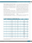
Table 1. Characteristics of the cohort of patients with del(5q) myelodysplastic syndromes.
Patient Age at first Sex n. diagnosis
1 66F
2 75F
3 78 F
4 68F
5 74 M
6 52 M
7 71 M
8 63 F
9 76 F
10 48 F
11 68 F
12 74 M
13 60 M
14 51F
15 45F
WHO diagnosis
Isolated del5q
Isolated del5q
Isolateddel5q
Isolated del5q
Isolated del5q
Isolated del5q
Isolated del5q
Isolated del5q
Isolated del5q
Isolated del5q
Isolated del5q
Isolated del5q
Isolated del5q
Isolated del5q
Isolated del5q
IPSS Samples
Int-1 BM, PB
Int-1 BM, PB
Int-1 BM,PB
Int-1 BM
Int-1 BM, PB
Int-1 BM, PB
Low BM, PB
Int-1 BM, PB
Low BM, PB
Int-1 BM
Int-1 BM
Int-1 BM
Int-1 BM
Int-1 BM
Int-1 BM
Months after 1st sample
0 12
0
16
Response at sample collection
Baseline PR
Baseline
No treatment
Best response
PR/TI
No
change/SD
0 Baseline TI 7 PR
0
15
0 30
0
Baseline
PD (morphology)
Baseline NA
Baseline
TI
CCR/TI
NA
CCR/TI
NA
CCR/TI
CCR/TI
PR/TI
TI
CCR/TI
CCR/TI
CCR/TI
7 SD
0 18
0
Baseline CR
Baseline
6 SD
0 42
0
30
0 12
0
6
0
6 CR
0 Baseline
12 CR
0 Baseline
6 CR
Baseline PR/CCR?
Baseline
Relapse
Baseline PD (RAEB-2)
Baseline
PD (RAEB-2)
Baseline
N: number; F: female; M: male; WHO: World Health Organization; Isolated del5q: myelodysplastic syndrome with isolated deletion of chromosome 5q; IPSS: International Prognostic Scoring System; Int-1: intermediate risk 1; BM; bone marrow; PB: peripheral blood; TI: transfusion independence; CR: complete remission; CCR: complete cytogenetic remission; PR: partial remission; SD: stable disease; PD: progressive disease; RAEB.-2: refractory anemia with excess blasts-2; NA: not available; Baseline: indicates sample taken before the initiation of lenalidomide treatment..
haematologica | 2019; 104(7)
1357
