
Page 168 - 2019_07 resto del Mondo-web
P. 168
J.R. Jones et al.
A
B
C
D
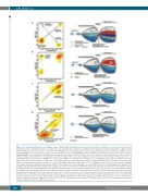
Figure 4. The evolutionary patterns seen leading to relapse. (A) Branching: branching evolution was the predominant mechanism seen and was characterized both by the gain and loss of mutational clusters at relapse. The cancer clonal fractions (CCF) for all coding mutations using kernel density estimation for a typical patient (left) is shown and reveals the presence of a new dominant PRDM1 (CCF 1.0) containing clone at relapse only (each dot represents a mutation). In addition, a clone containing CHD2 (CCF 0.91 presentation only) is lost at relapse while a clone containing NRAS remained dominant at presentation (CCF 1.0) and relapse (CCF 0.99). (Right) Illustration of the branching evolutionary process using the same patient. Prior to treatment, there are a number of competing sub-clones, but as a result of effective therapy, clonal extinction occurs leading to a genetic bottleneck. This leads to the emergence of a new clonal structure at relapse; in this case the loss of a dominant CHD2 clone, the gain of a PRDM1 clone, and a stable NRAS clone. In addition, the emergence of a new DDB1 mutation was seen within a minor clone with a CCF of 0.21. (B) Linear: linear evolution was seen in 20% of patients, characterized by the gain of mutations at relapse but no evidence of clonal loss. The KDE plot is displayed and shows the emergence of a new clonal PRDM1 mutation at relapse with a CCF of 1.0. (Right) Over time, successive generations of daughter cells acquire aberrations making them genetically distinct; in this example, we see the emergence of a new PRDM1 mutation. (C) Stable progression: KDE plot (left) showing a typical patient with stable progression, revealing a preserved clonal structure at both time points, with CCF values for all mutations remaining consistent at both time points. The CHD2 mutation was present within a dominant clone at presentation and relapse with a CCF of 0.83 and 0.87, respectively. Stable evolution was a characteristic of patients achieving a non-complete remission (non-CR), and in particular a partial remission (PR). These patients appeared to have a treatment resistant disease status and therefore the emergence of the same clonal structure was seen at relapse as had been seen at disease onset; in this case, with a CHD2 dominant clone at both time points (right). (D) Stable with loss was seen in one patient and kernel density estimation (right) revealed the presence of a predominantly preserved clonal structure at relapse with clusters containing TRAF3 and LTB present with similar CCF values at both points. There was evidence of the loss of a cluster of mutations at relapse, suggestive of clone loss (circled). The evolutionary process is shown. Treatment sensitive clone(s) are eliminated but the resistant clone(s) remain and lead to the relapse disease state.
1446
haematologica | 2019; 104(7)
