
Page 131 - 2019_07 resto del Mondo-web
P. 131
ERG deletion in BCP-ALL
Sty arrays (Affymetrix, Santa Clara, CA, USA) were available from our previous study.10
DUX4r-acute lymphoblastic leukemia classification, analysis of DUX4, RAG1 and RAG2 expression
DUX4r-ALL was classified by supervised hierarchical clustering of patients based on expression of DUX4r-ALL signature genes (the top 150 up-regulated and the top 150 down-regulated genes in DUX4r-ALL compared to non-DUX4r-ALL9). Gene expression profiling was performed by whole transcriptome sequencing (RNAseq) and/or on microarrays, as described previously.11,12 Expression of DUX4, RAG1 and RAG2 was analyzed using data from RNAseq. RAG1/RAG2 reads were aligned and counted using hg19 reference genome.13 DUX4 reads were mapped to the DUX4 reference sequence and counted as described previously.9 Read counts were normalized using library size factor computed using R package Deseq2.14
Analysis of ERGalt expression
Expression of “ERGalt a” and “ERGalt b”10 was analyzed using
RNAseq data. Reads containing the sequences specific for these ERGalt transcripts were counted and normalized by library size factors.
Amplicon sequencing
Libraries for the amplicon sequencing were prepared by one round multiplex PCR using FastStartTM High Fidelity PCR System (Roche, Basel, Switzerland). PCR primers used to amplify ERGdel spanning region and 1-2 control amplicons are listed in Online Supplementary Table S1 and their schematic position is shown in Figure 1. Sequencing was performed on an Ion Torrent PGM sequencer (Life Technologies, Carlsbad, CA, USA) using 400 bp chemistry according to the manufacturer's instructions (Life Technologies). Reads were successively mapped to a custom ref- erence using the Burrows-Wheeler Alignment (bwa) tool. First, reads were mapped to a custom reference comprising reference sequences for both control amplicons and reference sequences sur- rounding the five ERG 3’ breakpoint site clusters. Next, from all reads partially mapped on ERG 3’ breakpoint site clusters, the unmapped parts were exported and mapped against the reference sequence surrounding the common ERG 5’ breakpoint site. The unmapped parts of reads in between segments mapped to 3’and 5’
breakpoint site references were considered inserted non-templat- ed nucleotides (N-segment). Data analysis was the same as that used for V-(D)-J rearrangements of immunoglobulin/T-cell recep- tor genes. Identified ERGdel alleles were defined by the position of last non-deleted 5’ nucleotide, N-segment (inserted non-tem- plated nucleotides), type of utilized 3’ breakpoint site cluster, and position of the first non-deleted 3’ nucleotide. Sequencing setting (target coverage, choice of control amplicons) and the coverage achieved are shown in Online Supplementary Table S2.
Statistical analysis
The Mann-Whitney U test was used to compare numerical parameters in DUX4r-ALL stratified by ERGdel. The two-tailed Fisher exact probability test was used to compare frequencies. The Kaplan-Meier method was used to estimate survival rates, differ- ences were compared with the two-sided log-rank test. Event-free survival (EFS) was defined as the time from diagnosis to the date of last follow up in complete remission or to the first event. Events were resistance to therapy (non-response), relapse, secondary neo- plasm, or death from any cause. Failure to achieve remission due to early death or non-response was considered as events at time zero. Patients lost to follow up were censored at the time of their withdrawal.
Further details of the methods used are available in the Online Supplementary Appendix.
Results
Frequency of ERGdel in DUX4r- and non-DUX4r-acute lymphoblastic leukemia: performance of different ERGdel screening methods
We studied the presence of ERGdel in a cohort of 118 B- other ALL patients of whom 50 and 68 were assigned into the DUX4r or the non-DUX4r ALL subgroups, respective- ly, based on the presence of DUX4r-specific gene expres- sion signature9 and DUX4 gene rearrangements (see Online Supplementary Results).
Using SNP array, we found ERGdel in 16 of 47 (34%) DUX4r-ALL patients. In 12 of 16 positive patients, the SNP array findings corresponded to the most frequent type of ERGdel, targeted by PCR/AmpliSeq (IntERGdel), while a
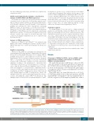
Figure 1. Schematic representation of the ERG gene, its transcript variants, and different types of ERGdel found in the present study. Black (gray) boxes represent exons of the gene (individual transcript variants). Accession numbers for reference sequences from NCBI Reference Sequence Database are shown. The most com- mon types of the deletion (IntERGdel) are shown in red, other types (ERGdel-diff) are shown in orange. Positions of primers used for the amplification of IntERGdel spanning region (green triangles) and 2 control amplicons (blue triangles) are shown. BP: breakpoint site.
haematologica | 2019; 104(7)
1409
