
Page 76 - 2019_06-Haematologica-web
P. 76
K.N. Smitheman et al.
A
BC
DE
FG
HI
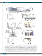
Figure 1. Inactivation of LSD1 inhibits the growth of acute myeloid leukemia (AML) cells by releasing myeloid differentiation block. (A) Percent maximum inhibition (bars) ± Standard Error and EC50 (circles) for 20 human AML cell lines treated with a titration of GSK2879552 for ten days. (B) Dose response of incorporation by BrdU Cell Proliferation Assay Kit (Cell Signaling Technology) of MOLM-13 cells treated with a titration of GSK-LSD1 for six days. (C) Fold caspase 3/7 activation (± Standard Error) relative to vehicle control of AML cell lines treated with 1000 nM GSK2879552 for 1-6 days or 100 nM bortezomib for three days. (D) Dose response of ITGAM (CD11b, left) and CD86 (right) gene expression on MOLM-13 cells treated with a titration of GSK2879552 for one day. (E) Representative flow cytometric histograms of CD11b protein expression on THP-1 cells (left) and CD86 protein expression on MOLM-13 cells (right) treated with GSK2879552 for one day. Concentrations of GSK2879552 from top to bottom are 0, 7, 47, and 3000 nM. (F) The change (treatment-vehicle) in the percentage of cells positive for CD86 (blue) and CD11b (red) protein expression (± Standard Error) for ten AML cell lines treated with 500 nM GSK2879552 (or 1000 nM GSK2879552*) for three days. †Significance (P<0.05) between dimethyl sulfoxide (DMSO) and GSK552 treatment. (G) Peak superoxide anion production (± Standard Error) of four AML cell lines treated with 1 μM GSK2879552 for 4, 7, 11, and 14 days. (H) Peak superoxide anion production of SKM-1 cells treated with a titration of GSK2879552 for seven days. (I) Dose responses of AML blast colony formation of 14 patient samples treated with a titration of GSK2879552. APL: acute promyelocytic leukemia.
1158
haematologica | 2019; 104(6)
