
Page 157 - 2019_06-Haematologica-web
P. 157
Glycoprotein V in ITP
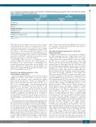
Table 1. Summary of autoantibody specificities detected in 343 of 1140 immune thrombocytopenia patients, either on the surface of the patient’s platelets (platelet-bound) or free in patient serum.
Glycoprotein specificity
GP IIb/IIIa only
GP Ib/IX only
GPVonly
GP IIb/IIIa plus GP Ib/IX
GP IIb/IIIa plus GP V
GP Ib/IX plus GP V
GP IIb/IIIa plus GP Ib/IX plus GP V
Total
*Rounding error.
ITP). This group was further assessed in order to ensure comparability of data. Results are summarized in Table 1. For patients with a positive test result for at least one gly- coprotein, the frequency of immunization against GP V was similar to the other glycoproteins: 242 out of 343 (70.6%) patients were positive for anti-GP IIb/IIIa, 232 out of 343 (67.6%) patients were positive for anti-GP Ib/IX, and 222 out of 343 (64.7%) patients were positive for anti- GP V (Kruskal-Wallis test; P=0.67) (Table 1).
Interestingly, there was also no difference in the amount of antibodies attached to GP V (antibody load), as determined by the optical density of the MAIPA assay between glycoproteins: mean values were 1.86 [95% con- fidence interval (CI): 1.49-2.23] for anti-GP IIb/IIIa, 1.63 (1.27-1.99) for anti-GP Ib/IX, and 1.82 (1.37-2.26) for anti- GP V; Kruskal-Wallis test, P=0.77.
Prevalence and binding properties of free autoantibodies against GP V
Sera from patients with any positive result in the direct MAIPA test (n=343) were further assessed by indirect MAIPA for the presence of free autoantibodies against these platelet glycoproteins. Results are summarized in Table 1. Free autoantibodies were detected in 45 out of 343 (13.1%) patient samples. The glycoprotein-specific distribution was GP IIb/IIIa (25 out of 45, 55.5%), GP Ib/IX (30 out of 45, 66.6%), and GP V (29 out of 45, 64.4%). Identified free autoantibody specificities matched the platelet-bound specificities from the same patient throughout. Addition of recombinant GP V to sera prior to testing completely blocked the detection of anti-GP V autoantibodies, but did not interfere with the detection of anti-GP IIb/IIIa or anti-GP Ib/IX autoantibodies (data not shown).
Serum IgG fractions from all 222 patients with platelet- bound anti-GP V were further analyzed by SPR (Figure 1); 88 out of 222 (39.6%) patients showed specific binding to the GP V flow cell. These 88 sera included all 29 identified as containing anti-GP V by indirect MAIPA. Further analy- sis demonstrated that these 29 sera with autoantibodies detected both in the indirect MAIPA and in SPR were of higher avidity (R700/R350=0.73±0.14; Wilcoxon rank test, P<0.001) (Figure 1B, top panel) than the 59 sera that gave positive signals in SPR, but were negative in the indirect MAIPA assay (R700/R350=0.32±0.13) (Figure 1B, bottom
Platelet-bound
autoantibodies N. of positive
samples %
71 20.7
30 8.8 10 2.9 20 5.8 10 2.9 61 17.8 141 41.1
Free autoantibodies
N. of positive samples
6
6 6 3 3 9 12
%
13.3
13.3 13.3 6.7 6.7 20.0 26.6 99.9*
343 100.0 45
panel). These results indicate that SPR has better sensitiv- ity compared to the gold standard MAIPA assay in detect- ing anti-GP V autoantibodies.
Autoantibody-triggered phagocytosis and in vivo platelet clearance
Anti-GP V autoantibodies were grouped according to their SPR binding profiles into a “high avidity” and a “low avidity” group. IgG fractions prepared from two high- avidity and two low-avidity anti-GP V antibody-contain- ing ITP sera were tested in a phagocytosis assay using CD14 positively-selected human macrophages from ITP spleens (Figure 2). One high- and one low-avidity GP V sera induced significant platelet uptake relative to normal human serum controls (P=0.003 and P=0.026, respective- ly). Of those positive, high- and low-avidity antibodies induced similar amounts of platelet uptake [mean phago- cytic index, 6.81 (range, 4.75-9.86) vs. 6.01 (range, 5.00- 6.98), respectively; P=0.954]. To further assess the biolog- ical effect of anti-GP V autoantibodies on platelet destruc- tion, the NOD/SCID mouse model was used. First, moab SW16 against human GP V was injected at two concentra- tions and the results verified against a murine monoclonal antibody (SZ21) specific for GPIIb/IIIa known to cause thrombocytopenia. SW16 induced similar clearance of human platelets from the murine circulation as SZ21 (mean platelet survival after 300 min, 16±5% vs. 8±8%; P=0.140) (Figure 3A). Platelet elimination was slower when SW16 was injected at a lower concentration (27±4%; P=0.018) (Figure 3A). Next, we analyzed IgG fractions isolated from ITP sera which contained anti-GP V autoantibodies only. Unexpectedly, anti-GP V reduced the survival of human platelets compared to control IgG regardless of their binding properties [median platelet sur- vival after 300 min, “high avidity”, 35% (range, 16-46%; P=0.029) and “low avidity”: 40% (range, 27-55%; P=0.025), respectively] (Figure 3B). After 24 h, only a few injected human platelet circulated in the presence of anti- GP V antibodies [median platelet survival after 1440 min, “high avidity”, 22% (range, 11-23%; P=0.0286) and “low avidity”, 20% (range, 13-24%; P=0.029) vs. 46% (range, 43-76%)] (Figure 3B). No difference in platelet elimination was observed between the two groups (P=0.229 and P=0.441, after 300 min and 1440 min, respectively). As expected, autoantibodies were generally less effective in
haematologica | 2019; 104(6)
1239
