
Page 78 - 2019_05-HaematologicaMondo-web
P. 78
E. Lombardi et al.
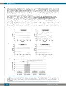
transverse velocity is a parameter that is correlated with the disturbed movement of a particle. The path of healthy RBC appeared to be more regular, uniform, and parallel to the flow (Figure 4A, Online Supplementary Video S2). Thus, healthy RBC crossed the field of view more rapidly than did sickle RBC (0.8-1.2 s versus 1.7-2 s, respectively; P<0.05). In the presence of FH, the trajectory of sickle RBC was regularized (Figure 4C), reducing their transverse velocity to 7.71 ± 6.7 mm/s (average decrease of 81.13% versus vehicle-treated sickle RBC). The same behavior appeared in sickle RBC treated only with the FH 19-20 domain (Figure 4D), with the transverse speed being 6.56 ± 5.6 mm/s (an average decrease of 83.94% versus vehicle- treated sickle RBC). When the particle kinematics were quantified (Figure 3E), we found that the absolute value of the instantaneous transverse speed of sickle RBC was
40.86 ± 27.6 mm/s, whereas that of healthy cells was only 5.64 ± 4.9 mm/s (a difference of 86.20%). When healthy cells were compared to either FH- or FH19-20-treated sick- le RBC, the absolute values for instantaneous transverse speed decreased significantly, reaching values similar to those observed for healthy RBC.
Anti-P-selectin and anti-Mac-1 antibodies prevent adhesion of sickle red blood cells to tumor necrosis factor-a-activated vascular endothelial surface
To better understand the binding proteins that may be important for adhesion of C3b/iC3b+ SCD red cells, we then evaluated the effects of anti-P-selectin or anti-Mac-1 antibodies on the adhesion of SCD to TNF-a-activated vascular endothelium. We chose these two molecules for the following reasons: (i) they are both modulated in
AB
CD
E
Figure 4. Factor H and its 19-20 fragment normalized the “stop-and-go” motion of sickle red blood cells. (A) Trajectory of three representative healthy (AA) red blood cells (RBC) in the field of view: each coordinate indicates their centroid at every consecutive frame (flow direction: x axis). (B) Trajectory of three representative sickle (SCD) RBC, showing the “stop-and-go” motion. (C) Trajectory of three representative sickle RBC treated with fator H (FH) (18 nM). (D) Trajectory of three representative sickle RBC treated with FH 19-20 segment (18 nM). (E) Absolute values for instantaneous transverse speed expressed as mean ± standard deviation (**P<0.01 ver- sus healthy RBC).
924
haematologica | 2019; 104(5)
