
Page 156 - 2019_04-Haematologica-web
P. 156
C. Recasens-Zorzo et al.
lymphocytes following JQ1 treatment,40 Figure 3B shows that CPI203 addition did not alter CXCR4 protein levels either in the presence or absence of IQS-01.01RS. Flow cytometry analysis further confirmed that surface levels of CXCR4 were unmodified by BET bromodomain inhi- bition (data not shown). To further ascertain the role of CXCR4 expression in this drug interaction, we estab- lished genetic depletion of CXCR4 in SUDHL-6 cells employing a CRISPR/Cas9 gene editing tool to generate CXCR4 knocked out (KO) cell lines, prior to treatment with CPI203, IQS-01.01RS and the combination of both agents. In parallel, and as a control of non-lymphomatous B malignancy, we used the recently described NALM6- CXCR4-KO cell line (kindly provided by Dr Jan Burger, Department of Leukemia, The University of Texas MD Anderson Cancer Center, Houston, TX, USA). As shown in Online Supplementary Figure S1, CXCR4-KO cell lines
completely lack CXCR4 molecules on their surface. The main difference between these two cell lines was the response to CPI203 as a single agent, which was substan- tially weaker in NALM6-CXCR4-KO cells. This may illustrate the recent finding that, in acute myeloid leukemia cells, CXCR4 signaling is involved in the regula- tion of MYC transcription mediated by the downregula- tion of the miRNA let-7a,41 while in DLBCL cells no vari- ation in MYC mRNA levels are observed following CXCR4 ligation (Online Supplementary Figure S2). Another explanation may come from the lower membrane expres- sion (about one log10) of the receptor observed in parental acute myeloid leukemia cells when compared to parental DLBCL cells, suggesting that NALM6 cells are less dependent on CXCR4 and less susceptible to CXCR4- dependent cellular stress than are SUDHL-6 cells. Most importantly, in the acute myeloid leukemia cell line the
AB
C
D
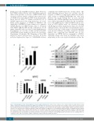
Figure 3. IQS-01.01RS synergizes with the BET bromodomain inhibitor CPI203 in vitro. (A) The relative antitumor effect of IQS-01.01RS (100 mM), CPI203 (0.5 mM) and the combination of both was determined by a MTT assay, after 48 h. The data shown are the mean results of the 13 DLBCL cell lines. (B) Cooperation between IQS-01.01RS and CPI203 in the inhibition of CXCR4 downstream signaling, as assessed by western blot analysis of p-AKT and MYC. SUDHL-6 and U2932 cells were starved for 2 h, and treated for 1 h with 100 mM IQS-01.01RS and/or CPI203 (0.5 mM) prior to a 1 min stimulation with 200 ng/mL recombinant CXCL12. α-tubulin was used as a loading control. (C) Relative MYC transcript levels in SUDHL-6 and U2932 cells upon 6 h treatment with 100 mM IQS-01.01RS, 0.5 mM CPI203 and the combination of both. Control untreated cells were used as a reference. (D) Time-dependent determination of MYC protein levels in SUDHL-6 cells treated with the translational blocker cycloheximide, as previously described,62 in the presence or absence of 100 mM IQS01.01-RS. β-actin was used as a loading control. CI: combination index; CHX: cycloheximide; COMBO: combination treatment with IQS-01.01RS and CPI203.
784
haematologica | 2019; 104(4)
