
Page 152 - 2019_04-Haematologica-web
P. 152
C. Recasens-Zorzo et al.
membranes (Immobilon-P; Millipore), or nitrocellulose mem- branes (Amersham Biosciences). Membranes were then incubat- ed with primary and secondary antibodies (Online Supplementary Materials and Methods) and visualized on a mini-LAS4000 device (Fujifilm) by enhanced chemiluminescence (ECL, Amersham Life Science).
Flow cytometry analysis
For the detection of surface CXCR4, DLBCL cells (2x10⁵) were stained with a phycoerythrin (PE)-labeled anti-CXCR4 or iso- type control antibody (BD Biosciences). For the CXCR4 occu- pancy assay, cells were pretreated for 1 h with IQS-01.01, AMD3100 (Sigma-Aldrich) or an anti-human CXCR4 control antibody, followed by sCXCR4 staining as above. For quantifi- cation of apoptosis, cells were labeled with Pacific Blue-conju- gated annexin V (Thermo Fisher). A total of 10,000 events were acquired and analyzed on an Attune acoustic focusing cytometer (Thermo Fisher). The mean fluorescence intensity ratio was cal- culated as the ratio between the median fluorescence intensity of the CXCR4-labeled sample and that of the isotype.
Chemotaxis assay
DLBCL cells (5x10⁶ cells/mL) were cultured for 1 h in culture medium not containing fetal bovine serum but supplemented with 0.5% bovine serum albumin (Sigma-Aldrich), in the pres- ence or absence of 100 mM AMD3100 or IQS-01.01RS, and ana- lyzed for CXCL12-dependent chemotaxis, as previously described.28 Values are referred to cells cultured without CXCL12.
Cell proliferation assay
DLBCL primary cultures and cell lines (5x10⁴ cells) were incu- bated for 24-48 h with IQS-01.01RS, AMD3100 and/or CPI203 (kindly provided by Constellation Pharmaceuticals) at the indi- cated doses. MTT (3-(4,5-dimethylthiazolyl-2)-2,5-diphenylte- trazolium bromide) reagent (Sigma-Aldrich) was added for 1–5 additional hours before spectrophotometric measurements. Each measurement was made in triplicate. Values are represent- ed using untreated control cells as the reference. Combination indexes were calculated using Calcusyn software version 2.0 (Biosoft). The interaction between two drugs was considered synergistic when the combination index was less than 0.8.
In vivo assays
Prior to efficacy testing, an acute toxicity assay was conducted
in NSG mice exposed to IQS-01.01RS salt or AMD3100. Animals (2 adult males and 2 adult females per dose) received a single administration of vehicle, IQS01.01-RS (per os) or plerix- afor (intraperitoneally) at doses ranging from 2 to 10 mg/kg, and were monitored during the first 4 h after administration, and daily for 2 weeks, for viability/mortality and vital parameters. This toxicity assay defined a maximum non-lethal dose of 5 mg/kg for AMD3100, while the maximum lethal dose of IQS- 01.01RS remained unreached.
For the systemic DLBCL model, 8-week old NSG female mice (n=12) were inoculated intravenously with 107 SUDHL6-GFP- Luc cells, randomly assigned into three equivalent cohorts, and treated daily with 5 mg/kg AMD3100 (intraperitoneally), 10 mg/kg IQS-01.01RS salt (per os) or vehicle. After 27 days, ani- mals were sacrificed and peripheral blood was collected by sub- mandibular puncture. Erythrocytes were lysed using ACK buffer (Quality Biological) and the percentage of SUDHL-6 cells was evaluated by detection of a GFP signal on an Attune cytometer. For the subcutaneous DLBCL model, SUDHL6-GFP-Luc cells were inoculated subcutaneously in 8-week old NSG female mice as previously.28,32 Animals were randomly assigned into four cohorts of four mice each and were given a twice daily dose of 1.5 mg/kg CPI203 (intraperitoneally), a daily dose of 2 mg/kg IQS-01.01RS (per os), both agents, or an equal volume of vehicle. Tumor engraftment was determined weekly following mice injection with 75 mg/kg D-luciferine (AnaSpec) and biolumines- cence imaging (BLI) using an Aequoria Luxiflux device equipped with an ORCA-ER camera (Hamamatsu). The bioluminescence imaging signal was quantified using Image J software. Tumor volume was measured by external calipers twice a week, up to 28 days. Animals were then sacrificed and tumors were harvest- ed and weighed. Animals were handled following protocols approved by the Animal Ethics Committee of the University of Barcelona (protocol #154/16).
Statisticalanalysis
A Wilcoxon rank-sum test, Fisher exact test, Spearman rank correlation or t-test was used to examine the statistical signifi- cance of associations between clinico-pathological data and CXCL12-CXCR4 positivity or cytokine expression level, as appropriate. Survival curves were estimated using the Kaplan- Meier method. A log-rank test was used to compare survival curves between groups. For in vitro and in vivo functional assays, unpaired and paired t-tests were employed to obtain the statisti- cal significance. The Benajmini-Hochberg method was used to adjust P-values for multiple testing. Results were considered sta- tistically significant when the P value was less than 0.05.
Results
CXCL12 expression correlates with microvascular density and confers an unfavorable prognosis
to patients with diffuse large B-cell lymphoma
Gene expression profiling studies have highlighted the prognostic value of the microenvironment and compo- nents of several immune regulatory pathways in DLBCL.4 To characterize which cytokines may have a significant impact on the pathogenesis of DLBCL, we performed a fluorescence-based cytokine antibody array using frozen tissue from 12 DLBCL cases, out of an initial cohort of 52 patients (Online Supplementary Table S1), and correlated expression data with clinico-biological characteristics of
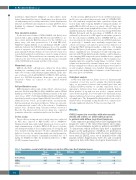
Table 1. Top cytokines associated with high microvessel density in diffuse large B-cell lymphoma biopsies.
Cytokine
SDF1α/CXCL12 IGFBP2
IGFBP1
MVD: microvessel density.
Mean (MVD=low)
5.207
4.467
5.085
Mean (MVD=high)
5.637
4.946
5.601
Difference
0.430
0.479
0.516
t statistic
-3.758
-3.744
-3.666
P
0.000
0.000
0.001
Adjusted P
0.032
0.032
0.032
780
haematologica | 2019; 104(4)
