
Page 24 - 2019_03-Haematologica-web
P. 24
430
Perspective Article
continued from the previous page
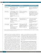
Model
Static conditions
Acellular synthetic microvascular model
Cellular synthetic microvascular model
In vivo microvascular model (large animal)
In vivo microvascular model (small animal)
Protocol
• Soft lithography with vessel diameters of 5-70 mm in PDMS with no cell lining
• RBCs perfused through system at different concentrations
- 0.4 haematocrit (30)
• Soft lithography with vessel diameters
of 50-200 mm in PDMS (9) or collagen (31)
• Channels lined with HUVECs (9, 31) • RBCs perfused at 10-20% (v/v) RBCs
with shear stress up to 17 dyn/cm2 (9, 31) • Model can be stretched to simulate
respiratory forces (9)
• Perivascular cells can be added to collagen
matrix (31)
• Microvasculature assessed with - Sublingual sidestream dark field
Outcome measures
• Perfusion rate for RBCs stored for different periods (30)
• RBC adhesion, endothelial marker expression, confocal microscopy (31)
• Endothelial damage (9)
• Microcirculatory flow index, microvascular blood flow, capillary-venous
hemoglobin oxygen saturation (46)
• RBC velocity, vessel diameter, FCD, after stored PRBC transfusion (47) • FCD, RBC velocity, vessel diameter,
O2 distribution after PRBC transfusion (50) • RBC adhesion in capillaries (48)
• RBC velocity, vessel diameter (49)
Research output and clinical impact
• Perfusion rate for stored RBCs was 19-30% lower than for fresh RBCs. Washing stored RBCs in saline improved perfusion rate
by 41% (30)
• Low hemodynamic shear stress due
to altered microcirculatory flow may predispose HUVECs to necroptosis (9)
• Cyclic stretching of microvessels (similar to breathing movements or mechanical ventilation) may increase susceptibility of
HUVECs to transfused RBCs (9)
• Isotonic or hypertonic colloidal fluids adequately restored sublingual microcirculatory blood flow and
flow quality (46)
• Gelatin + hydroxyethyl starch improved microvascular hemoglobin oxygen saturation (46)
• Fresh PRBCs more effective at relieving microcirculatory hypoxia compared to stored PRBCs in rat cremaster flap
model (47)
• IgG-mediated HTRs induced acute
vaso-occlusive crisis in the mice cremaster model. CXCR2 blockage prevented HTR-induced vasoocclusive crisis (49)
imaging (46)
- Laser Doppler flowmeter (46)
- Tissue reflectance spectrophotometer (46)
• Animals used
- Domestic pig (46)
• Animals transfused with blood product and then assessed using intravital microscopy
• Animals used
- Rat: cremaster flap (47), extensor
digitorum longus muscle (48) - Mice cremaster flap (49)
- Hamster: dorsal skin flap (50)
BC: buffy coat; E. coli: Escherichia coli; FCD: functional capillary density; HTR: hemolytic transfusion reaction; HUVECs: human umbilical vin endothelial cells; LPS: lipopolysaccharide; PC: platelet concentrate; PDMS: polydimethylsiloxane; PMPs: platelet microparticles; PRBC: packed red blood cells; PRP: platelet-rich plasma; RBC: red blood cell; RMP: red cell microparticle; ROS: reactive oxygen species; TNF-α: tumour necrosis factor--alpha.
alignment.6 Lastly, shear stress modifies endothelial inter- actions with blood cells. For example, monocytes per- fused over endothelium activated by tumor necrosis fac- tor-α expressed more tissue factor and CD11b compared to monocytes co-incubated with activated endothelium under static conditions.9
It follows that intravascular transfusion events are affected by blood product-recipient blood interactions and blood mixture-endothelium interactions under flow conditions. Simulation of the post-transfusion intravascu- lar mileau in vitro requires recipient whole blood, blood product, endothelium and perfusate flow. To date, the majority of models used to test transfusion effects have involved static models in which blood products are co- incubated with specific cells such as neutrophils,1,17-21 macrophages,3 platelets22 or endothelial cells23 to examine interactions between two cell types. While blood co-incu- bation experiments enable the investigation of multiple cellular interactions,2,24 they do not recapitulate the fre- quency and type of cellular interactions that occur under endogenous flow conditions. In vivo models are the “gold standard” for capturing the sum of intravascular interac-
tions that occur after transfusion of stored, packed RBCs. However, such models are limited by increased variabili- ty (requiring larger sample sizes), reduced capacity to iso- late parameters and longer set-up times. Endothelialized in vitro flow models circumvent these limitations by using cell culture methods, packed RBCs and fresh whole blood, which are easier to acquire with fewer associated ethical considerations, shorter model development time and lower cost.
What can we do with current manufacturing technology?
The potential methods to investigate blood mixture- endothelial interactions and current research outputs from these models are summarized in Table 1. Flow mod- els can be divided into planar, microvascular and macrovascular depending on the arrangement of endothelial and blood cells. For planar models, endothe- lial cells were cultured on coverslips and subjected to var- ious shear stresses generated by laminar flow.25-27 These models were used to study blood-endothelial adhesion under macrovascular flow conditions with shear stresses of 0.3-10 dyn/cm2.25-27 Notably, these models do not reca-
haematologica | 2019; 104(3)
