
Page 101 - 2019_03-Haematologica-web
P. 101
Atypical TG2 expression activates the NF-κB pathway
A
B
C
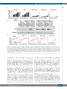
Figure 1. Tissue transglutaminase (TG2) accelerates antimicrobial ability of differentiating NB4 cell lines. (A) Relative mRNA expression of TG2 in NB4-wild-type (WT) treated with 1 μM all-trans retinoic acid (ATRA), virus control TG2-C, shRNA-silenced TG2-KD, hetero-allelic TG2-ha, and TALEN-TG2 knocked-out (KO) cells meas- ured at the indicated days by real-time Q-PCR and normalized to glyceraldehyde 3-phosphate dehydrogenase (GAPDH) mRNA expression (n=3). (B) Representative Western blot showing TG2 protein expression levels upon ATRA treatment over 11 days (n=3). (C) NB4 cell lines undergoing differentiation characterized by ability to reduce nitro-blue-tetrazolium (NBT). The assays were performed at the indicated time points in triplicates. Percentage of the NBT-positivity expressed as mean %±Standard Deviation for 3 parallel experiments. Statistical analysis was performed via two-way analysis of variance (ANOVA; Bonferroni post-hoc test; *P<0.05, **P<0.01, ***P<0.005 ****P<0.001).
differentiation on ATRA-treated NB4 cell lines, showed that while the expression of L-selectin, CD11b, and CD11c increased significantly from day 0 to day 3, subse- quently remaining almost unaltered regarding cell surface positivity (Online Supplementary Figure S5), the mean fluo- rescence intensity (MFI) of the cells increased during the 11-day treatment. ATRA-induced differentiation is associ- ated with increasing TG2 expression parallel to the pro- gression of the differentiation of NB4 cell lines. Neither knockdown of TG2 expression nor use of the NB4 TG2- KO cell line changed the cell-surface expression of L- selectin, CD11b, and CD11c in ATRA-induced differenti- ation (Figure 2A-C). In addition, PMA did not stimulate cell-surface expression of CD11b over basal expression (Figure 2B). Notably, some non-activated adhesion recep- tors were observed on the cell surface, while others were restricted to cytoplasmic granules, like CD11b.26 To gain insight into the activation states of ATRA-treated NB4 cell lines, we used CBRM1/5 mAb, which is specific for high- affinity CD11b and does not recognize CD11b on resting myeloid cells, and examined the cell-surface expression and affinity status of CD11b. We found that ATRA not
only enhanced the cell-surface expression of CD11b but also induced its high-affinity state (Figure 2B). Assuming that the measured surface expression levels of CD11b might be the “basal expression levels” on NB4 cell lines, we determined the PMA-stimulated CD11b cell-surface expression levels by CBRM1/5, revealing that PMA can- not further increase cell-surface expression of CD11b inte- grin (Figure 2B). Antibody anti-CD11c Clone 3.9 binds to CD11c in an activation-dependent manner and is specific to the I domain of CD11c.27 Anti-CD11c antibody revealed gradually increasing expression of activated CD11c integrin receptors on ATRA-treated NB4 cell lines, which could not be further enhanced by PMA (Figure 2C).
TG2 drives the respiratory burst of NB4 cell lines
Some phagocytes and neutrophil granulocytes can gen- erate large amounts of reactive oxidants in response to particulate or soluble inflammatory stimuli. We had previ- ously found that neutrophils of TG2-KO mice showed lower expression of both GP91PHOX mRNA and protein with less ROS production compared to wild-type mice.20 The TG2-dependence of PMA-induced ROS capacity was
haematologica | 2019; 104(3)
507
