
Page 208 - 2019_02-Haematologica-web
P. 208
M. Rijkers et al.
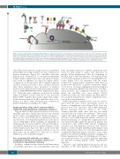
Figure 8. Proposed mechanism of anti-HLA antibody induced complement deposition. Upon binding of anti-HLA antibodies to HLA molecules on platelets, C1q can bind to HLA-bound IgGs. This leads to initiation of the classical complement pathway which results in C4b and C3b deposition on the platelet surface. Eventually this leads to the formation of a MAC which promotes the influx of Ca2+. Elevated intra-platelet Ca2+ levels induce platelet activation as measured by CD62P exposure. Anti- HLA antibody-induced complement activation can be inhibited using anti-C1q blocking antibody or Eculizumab at the C1q of C5 level, respectively. Via a separate mechanism binding of HLA antibodies to HLA molecules can cross-link with FcγRIIa and induce platelet activation independent of the complement pathway.
these HLA mAbs induced a large increase in membrane permeability providing evidence for pore formation in platelet membranes (Figure 5C). Anti-HLA antibodies induced pore formation in a concentration-dependent manner (Online Supplementary Figure S2) and pore forma- tion could be inhibited by anti-C1q (Figure 5D-E). Comparison of C3b deposition, C5b-9 complex formation and pore formation revealed a clear correlation between these three parameters upon incubation of platelets derived of two different donors with increasing concentra- tions of SN607D8/SN230G6 (Online Supplementary Figure S2). Under the same conditions, an increase in calcium influx in platelets loaded with fluo-4 was measured (Figure 5F-H). Together, these results suggest that comple- ment activation induced by HLA antibodies leads to for- mation of a MAC, with subsequent pore formation in platelet membranes resulting in Ca2+ influx.
Immunoglobulins (IVIg) and C1 esterase inhibitor inhibit HLA antibody-induced complement activation
FcγRIIa-dependent α-granule release was also investi- gated with the same donors and platelet donors, in absence of complement source (Figure 7D-F). Some sera induced both C3b deposition and FcγRIIa-dependent platelet activation, however, similar to the results obtained with HLA mAbs, there were also sera which activated only one of these two pathways. These results suggest that HLA antibodies may induce complement activation and FcγRIIa-dependent platelet activation via distinct mechanisms. To confirm that complement activa- tion induced by HLA antibodies present in patients sera correspond to that of HLA mAbs, platelet complement activation and Syk-dependent activation were blocked. Similar to results obtained with HLA mAbs (Figure 4 and Online Supplementary Figure S1), both Syk-dependent and complement-dependent platelet activation occurs when platelets are incubated with HLA antibody containing sera (Online Supplementary Figure S5). Furthermore, C3b depo- sition induced by these sera could be inhibited employing the IgG-Fc:Fc blocking peptide (Online Supplementary Figure S6).
IVIg can inhibit FcγRIIa-dependent platelet activation by anti-HLA antibodies.8 Similar effects have been observed in the context of heparin-induced thrombocytopenia.28,29 Here, we also tested if IVIg affects complement activation and observed a dose-dependent inhibition of C3b deposi- tion by IVIg (Figure 6A-B). Similarly, by inhibiting C1 employing the C1 esterase inhibitor, C3b and C4b deposi- tion induced by HLA mAbs was inhibited (Figure C-E and Online Supplementary Figure S1). These results show that complement activation on platelets induced in vitro by HLA mAbs can be inhibited by IVIg and C1 esterase inhibitor in a dose dependent manner.
Sera containing HLA antibodies can induce complement activation on platelets, which is not correlated to Syk-mediated activation
In order to confirm our data obtained with human mon- oclonal HLA antibodies, sera containing HLA antibodies
The level of IgG binding differed among sera and also depended on the donor platelets used (Figure 7G and
from 12 patients refractory to platelet transfusions were tested for their ability to induce C3b deposition on platelets (Online Supplementary Table S2). Depending on the HLA type of the donor platelets, C3b deposition was observed upon incubation with sera containing HLA anti- bodies (Figure 7A-C). In agreement with the results obtained for human mAbs, incubation with anti-C1q blocked C3b deposition on platelet membranes (Figure 7A-C). In case of strong complement activation, 25 mg/ml anti-C1q only partly inhibited C3b deposition. C3b depo- sition, however, was completely blocked by anti-C1q when the anti-HLA sera were diluted (Online Supplementary Figure S4). This confirms that HLA antibod- ies, as present in patient sera, induce complement activa- tion via the classical pathway.
412
haematologica | 2019; 104(2)
