
Page 130 - 2019_02-Haematologica-web
P. 130
B. Schuhmacher et al.
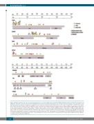
Figure 2. Mutation patterns in the most recurrently mutated genes in nodular lymphocyte-predominant Hodgkin lymphoma and T-cell/histiocyte-rich large B-cell lymphoma. Schematic overview of the mutational distribution in the most recurrently mutated genes. For each gene missense (triangles), stop gain (spheres) and splice site (squares) mutations are mapped to coding exons (top) and protein domains (bottom). Mutations are color-coded according to the occurrence in the groups (blue: NLPHL A/B; green: NLPHL C/D/E; red: THRLBCL). Amino acid positions of protein domains are adopted from the Uniprot database (www.uniprot.org) and refer to the canonical sequences (JUNB: P17275, DUSP2: Q05923, SGK1: O00141, SOCS1: O15524, CREBBP: Q92793, FN1: P02751, TRRAP: Q9Y4A5). bZIP: basic leucine zipper motif; DsPc: dual specific phosphatase, catalytic domain; Pkinase: protein kinase domain; AGC-kinase C: AGC-kinase C-terminal domain; Rhod: rho- danese; SH2: Src homology 2 domain; KIR: kinase inhibitory region; CH1/2/3: cysteine/hystidine-rich region; KIX: kinase inducible domain; BROMO: bromodomain; HAT: histone acetyltransferase domain; Q: poly glutamine stretch; FN: fibronectin; TP53: tumor suppressor p53 binding site; FAT: FRAP-ATM-TRRAP domain; FATC: FAT C-terminal; PI3K/PI4K: phosphatidylinositol 3-kinase/ phosphatidylinositol 4-kinase domain; NLPHL: nodular lymphocyte-predominant Hodgkin lymphoma; THRLBCL: T-cell/histiocyte-rich large B-cell lymphoma.
334
haematologica | 2019; 104(2)
