
Page 37 - 2019_01-Haematologica-web
P. 37
CNS prophylaxis in DLBCL
include the bone marrow, paranasal sinus, orbit, pericardi- um, ovary, uterus, and breast.37-41 Involvement of paranasal sinuses was identified as a significant risk in the pre-ritux- imab era, but recent data have demonstrated that the risk of CNS events is not increased in the era of rituximab- based therapy, and there is not a clear role for either intrathecal prophylaxis or consolidative radiation therapy in these patients.42 Kidney and adrenal involvement had previously been identified as high-risk locations, and are now included in the CNS-IPI, as previously discussed.43,44
Biological risk factors
As our understanding of the molecular pathogenesis of DLBCL has advanced, specific biological features have become increasingly important predictors of CNS risk. DLBCL harboring a MYC rearrangement has been associ- ated with an increased risk of CNS recurrence and an infe- rior overall survival relative to other forms of DLBCL.45,46 A MYC rearrangement rarely occurs as a sole genetic abnor- mality, however, and the dominant prognostic impact appears to be conferred by the rearrangement in concert with additional genetic aberrations.45,46 High-grade B-cell lymphoma with translocations of MYC and BCL2 and/or BCL6, also known as double- or triple-hit lymphoma, rep- resents about 5% of all newly diagnosed large B-cell lym- phomas and carries a poor prognosis with a median over- all survival of less than 2 years.47,48 CNS involvement is common at either diagnosis or relapse, and has been reported in as many as 50% of affected patients.48-50 Compared to routine DLBCL therapy, more aggressive ini- tial chemoimmunotherapy regimens such as dose-adjust- ed EPOCH-R (etoposide, prednisolone, vincristine, cyclophosphamide, doxorubicin plus rituximab) are typi- cally employed, along with intrathecal CNS prophylaxis. This is based primarily on retrospective analyses which have demonstrated improved progression-free survival with more aggressive induction regimens and have sug- gested improved survival for patients receiving intrathecal therapy in this disease in which CNS recurrences com- monly involve the leptomeningeal compartment.47
Another area of interest has been DLBCL with immuno- histochemically detectable expression of MYC and BCL2 without associated translocations, otherwise called dou- ble-expressing lymphoma. Dual protein expression is sig- nificantly more common than double-hit lymphoma, occurring in approximately 30% of cases of DLBCL.51,52 A retrospective analysis of double-expressing lymphoma by the BCCA found a CNS recurrence risk of approximately 9%, compared to only 2% in non-double-expressing cases of DLBCL.51 This risk was modified, however, based on cell of origin, and by risk stratification according to the CNS-IPI score. The risk of CNS events in double-express- ing lymphoma appears limited to the ABC-like subset of DLBCL in which the CNS relapse risk is approximately 15%, while there is no apparent increased risk in double- expressing germinal center B-cell (GCB)-like disease. Risk also appears confined to the intermediate- and high-risk CNS-IPI patients in whom the CNS relapse rate approxi- mates 12% and 22%, respectively, without any increased risk among low-risk CNS-IPI patients with double- expressing lymphoma. These analyses reinforce the com- plexity of assigning risk of CNS relapse in DLBCL, which warrants attention to clinical, histopathological, and molecular factors for optimal risk estimation.
Novel methods for risk stratification are being devel-
oped. The use of pretreatment positron emission tomo- graphy to predict CNS relapse has been proposed, with elevated total lesion glycolysis found to be predictive of an increased risk of CNS relapse on multivariable analysis.53 Additional biomarkers emerging from pathological analy- ses have been utilized to stratify risk, including ITGA10, CXCR5 and nuclear PTEN.54,55 Further research in these areas in concert with existing biomarkers and clinical risk stratification tools should be performed to determine the clinical utility of these studies before they are incorporat- ed into routine clinical care.
Baseline central nervous system evaluation
CNS recurrence of DLBCL typically occurs early in the disease course, either during systemic treatment or with- in several months of completing treatment.8,28 This sug- gests that subclinical involvement of the CNS by DLBCL is likely present at the time of diagnosis in such cases. Early identification of patients with CNS involvement is crucial, as the treatment and prognosis may be signifi- cantly altered based on this knowledge. As such, evalua- tion of the CNS via CSF analysis and/or neuroimaging should be considered in patients with high-risk features or neurological symptoms.
CSF analysis consists of conventional cytology and flow cytometry. Cytology alone has low sensitivity for CNS disease at less than 60% for leptomeningeal disease and virtually no ability to detect parenchymal lym- phoma.56 The addition of flow cytometry significantly increases sensitivity for detection of occult CNS involve- ment, which can be found in approximately 10% of high-risk patients and is associated with a high rate of subsequent CNS progression and poor overall survival.11- 15 An important caveat is that these data include patients predominantly treated in the pre-rituximab era, at a time when most CNS relapses involved the leptomeningeal compartment.28,57,58 Since the introduction of rituximab- based chemoimmunotherapy, the incidence of CNS
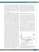
Figure 1. Kaplan-Meier curve depicting risk of central nervous system relapse based on the Central Nervous System International Prognostic Index score. DSHNHL: German High Grade Lymphoma Study Group cohort, BCCA: British Columbia Cancer Agency cohort. Schmitz, et al. J Clin Oncol 2016.18
haematologica | 2019; 104(1)
27
