
Page 210 - 2019_01-Haematologica-web
P. 210
A. Laghmouchi et al.
AB
C
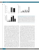
Figure 1. Reactivities within allo-reactive T-cell responses provoked by stimulation with HLA-DP-mismatched dendritic cells. (A) Frequencies (%) of CD137+ cells in the CD4+ T-cell population at 36 h after restimulation [white bars = responder cells not restimulated (NR), gray bars = responder cells stimulated with autologous CD14-derived dendritic cells (autoDC), black bars = responder cells restimulated with allogeneic CD14-derived dendritic cells (alloDC)]; representative example. (B) Frequencies (%) of CD137+ T cells in the CD4+ T- cell populations of responses 1-5 at 36 h after restimulation of the responder cells that underwent a previous depletion step of autoDC reactive T cells (white bars = NR responder cells, gray bars = autoDC-stimulated responder cells, black bars = alloDC-restimulated responder cells). (C) Reactivity of the expanded T-cell clones from the allo-reactive T-cell responses 1-5 (n=107, 158, 540, 581, and 293, respectively) (white bars = non-reactive T- cell clones, gray bars = autoDC reactive T-cell clones, black bars = alloDC reactive T-cell clones).
stimulation assay against third-party DC and EBV-LCL expressing the mismatched HLA-DP alleles (Online Supplementary Figure S3A) that were targeted in the immune responses, using autoEBV-LCL, autoDC and alloDC as controls. The majority (48-99%, median 73%, black bars in Figure 2A) of the allo-reactive T-cell clones showed HLA-DP-restricted reactivity demonstrated by recognition of alloDC and third-party EBV-LCL expressing the targeted mismatched HLA-DP allele (response 4, as a representative example, is shown in Online Supplementary Figure S3B). A representative HLA-DPB1*03:01-restricted T-cell clone is shown that only recognizes DC and EBV- LCL, but no other hematopoietic cells (CD14+ and PHA- blasts) (Figure 2B). However, a significant proportion (1- 52%, median 7%, gray bars in Figure 2A) of the allo-reac- tive T-cell clones recognized alloDC, but showed no reac- tivity against the third-party EBV-LCL expressing the tar- geted mismatched HLA-DP allele (response 4, as a repre- sentative example, is shown in Online Supplementary Figure S3C). Selected T-cell clones from this group were tested against third-party DC and showed recognition of these stimulator cells by production of interferon-γ (response 4, as a representative example, is shown in Online Supplementary Figure S3D). A representative HLA- DPB1*03:01-restricted T-cell clone that recognizes only DC and no other hematopoietic cells is shown (Figure 2C). To confirm the specific HLA-DP restriction, T-cell clones were tested against K562 cell lines transduced with specif- ic HLA-DP alleles or with empty vector (mock) (Online Supplementary Figure S4A). The T-cell clones only produced interferon-γ against the K562 transduced with the target HLA-DP allele (HLA-DPB1*03:01; representative T-cell clones, response 4 in Online Supplementary Figure S4B).
To analyze whether the differential cell-recognition pat- terns of the allo-HLA-DP reactive T-cell clones is caused by recognition of a polymorphic antigen (minor histocom- patibility antigen), a larger panel of DC and EBV-LCL was used as stimulator cells. The allo-reactive T-cell clones that showed HLA-DP-restricted reactivity against both DC and EBV-LCL manifested HLA-DP-restricted recognition of all DC (Figure 2D; representative HLA-DPB1*03:01- restricted clone) and EBV-LCL (Figure 2F; representative HLA-DPB1*03:01-restricted clone) expressing the target HLA-DP allele, indicating that most likely a monomorphic peptide is recognized. The allo-reactive T-cell clones that showed HLA-DP-restricted reactivity against DC but not against EBV-LCL showed HLA-DP-restricted recognition of the extended panel of DC (Figure 2E; representative HLA-DPB1*03:01-restricted clone) but not of the extended panel of EBV-LCL (Figure 2G; representative HLA- DPB1*03:01-restricted clone). Furthermore, the restricted recognition of DC but not of EBV-LCL from the same indi- vidual (e.g. DC and EBV-LCL from individuals 2, 3, 9 and 10 in Figure 2E,G) indicates that this restricted recognition profile is most likely caused by recognition of a cell-lin- eage-specific monomorphic peptide in the context of the allo-HLA-DPB1*03:01molecule.
In response 5, with an additional HLA-DQB1 mismatch, 38% of the allo-reactive clones did not show HLA-DP- restricted reactivity against EBV-LCL expressing the mis- matched HLA-DP molecules. Testing of these clones against a panel of DC and EBV-LCL expressing the mis- matched HLA-DQB1*06:03 allele revealed that 33% of the allo-reactive clones exerted HLA-DQB1-restricted reactiv- ity against DC and EBV-LCL, and 5% against DC, but not against EBV-LCL (bar 5b in Figure 2A; reactivity of repre-
200
haematologica | 2019; 104(1)
