
Page 19 - 2018_12-Haematologica-web
P. 19
Editorials
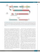
Figure 1. BCR-ABL rearrangement structure. The BCR gene contains three primary breakpoint clusters, the “minor” (m-bcr), “major” (M-bcr), and “micro” (μ-bcr). Gene rearrangement at the M-bcr site results in either of two p210 fusion chimeric mRNAs, composed of BCR exons 1-13 or exons 1-14 (orange and green) fused to ABL exons 2-11 (red). RT-PCR of BCR-ABL, amplifying only the exons, yields an amplicon product of ~200-300 bp. However, the genomic breakpoints in BCR and ABL are dispersed over intervals of 3.0 kb and 150 kb, respectively, making straightforward amplification of the DNA breakpoint impossible. The relative scale of the RNA and theoretical DNA product using the same set of BCR and ABL primers is shown at the bottom of the figure. Thus, for successful DNA amplification, multiple BCR and ABL primers must be used until a successful combination successfully amplifies the breakpoint. After this, sequencing is performed to identify sequences for patient-specific primers and probes.
believe that a series of environmental insults would affect both target and housekeeping genes in the same way. In addition, there are certainly differences in BCR-ABL levels from patient to patient, if not from cell to cell in the same patient. A DNA-based assay would be a more accurate measure of defining the approximate number of actual CML cells in any given patient. (Note that in rare cases a patient can harbor more than one copy of BCR-ABL, but this is typically only in cases of advanced phase disease.)
Amplification of the BCR-ABL DNA is difficult, as the potential span of breakpoints within the BCR and ABL genes is vast (Figure 1), as opposed to the limited base pair distance once the chimeric BCR-ABL mRNA is assembled.9,10 To perform the DNA-based assay for BCR- ABL, an initial PCR uses multiple primer sets to first iden- tify the possible DNA breakpoint. Once the sequence of the breakpoint is identified, patient-specific primers are constructed to make a very sensitive assay. Since the patient-specific PCR will have different kinetics from patient to patient, the drop in the disease burden must be measured against the patient's diagnostic disease burden value. This is a very similar concept to following MRD in patients with acute lymphoblastic leukemia, where patient-specific IgVDJ or TCR rearrangements must be amplified with consensus primers, then patient-specific primers and probes developed for each unique assay. [This complexity led to the development of sensitive flow cytometry and next generation sequencing (NGS) meth- ods, the latter discussed below.]
The potential value of a sensitive DNA test is at least 2- fold. First, one could study the differences in RNA versus DNA load, correlating this with disease response. Secondly, a DNA assay would allow the detection of CML in cases in which the RNA assay of BCR-ABL is undetectable. This would be especially interesting in the case of discontinuation in CML, where some patients with a prolonged deep molecular response stop TKI treat- ment and do not relapse.11,12 Are those cases that quickly relapse after discontinuation simply those in whom the disease is at a higher burden, though undetectable by RNA assays? Could patients who have no MRD by RNA or DNA assays be the lucky patients for whom discontin- uation will be successful?13
In this issue of the Journal, the Adelaide group expands on their previous studies of DNA-based BCR-ABL detec- tion and show the potential of this assay to probe basic disease and clinical issues.14 They studied 59 patients with 516 samples on which RNA and DNA assessments of dis- ease burden were performed. Several important findings were found.
First, they found that, early in disease treatment, the number of copies of RNA was generally higher than the DNA (roughly 2-fold) whereas after around six months, RNA and DNA levels were fairly similar. The biological reason for this is unclear. However, the kinetic decay of BCR-ABL with TKI therapy shows a multi-order decay, with an initial decline, followed by a slower decay. These two findings combined suggest that, at diagnosis, a pop-
haematologica | 2018; 103(12)
1943
