
Page 84 - 2018_10-Haematologica-web
P. 84
W. Zhang et al.
patient, previously treated with quizartinib and idarubicin with cyatarabine plus crenolanib, who achieved CRi with molecular CR for both FLT3-ITD and FLT3-D835 with selinexor plus sorafenib; (iii) a FLT3-mutated patient pre- viously treated with crenolanib, sorafenib and allogeneic stem cell transplantation who achieved CRi without molecular CR with selinexor plus sorafenib; and (iv) two FLT3-ITD and FLT3-D835 dual mutated patients who had received prior sorafenib therapy and achieved PR + >50% blast reduction with selinexor plus sorafenib (Daver et al., ASH annual meeting, 2017, Abstract #1344). We have no information regarding differentiation induction in these patients at this time.
Discussion
A previous study using the XPO1 inhibitor KPT-185 demonstrated strong post-transcriptional downregulation of total FLT3 protein expression in AML cell lines and pri- mary AML samples, which was associated with anti- leukemia efficacy.21 We confirmed the suppression of phospho-FLT3 and accompanying downregulation of total FLT3 protein in human MOLM13 and MV4-11 cells after 24 h of selinexor treatment (Online Supplementary Figure S9). Unexpectedly, however, we observed marked upreg- ulation of phospho-FLT3 in murine AML cells, including those harboring ITD, ITD plus D835Y or Y842C dual mutations, after exposure to the same concentrations of selinexor. Furthermore, total FLT3 protein and mRNA lev- els were upregulated in addition to the upregulation of FLT3 downstream phospho-ERK and -AKT levels in cells with these mutations, but not in the ITD plus D835Y mutants (Figure 1E, Online Supplementary Figure S4), sug- gesting that a possible transcriptional mechanism was
involved in FLT3 upregulation in these murine cells. The modulation profiling suggests that selinexor-induced apoptosis is independent of the suppression of FLT3 and its downstream pathways. In fact, cells with the dual Baf3/ITD+D835 mutation showed greater sensitivity to selinexor-induced apoptosis compared with other dual- mutated cells, Baf3/ITD+Y842 and Baf3/ITD+F691 (EC50 values were 0.2 and 0.3 mM versus 0.56 and 0.6 mM, respec- tively, Baf3/ITD+D835H and Baf3/ITD+D835Y versus Baf3/ITD+Y842 and Baf3/ITD+F691 cells), and sup- pressed phospho-ERK and –AKT, but upregulated phos- pho-FLT3 levels. These results strongly support that selinexor induces apoptosis in an FLT3-independent man- ner in FLT3-mutated AML cells. In addition, we observed upregulation of XPO1 cargo proteins p53, p21 and p27, as well as downregulation of pro-survival Mcl-1 after 24 h of selinexor treatment in both human and murine FLT3- mutated AML cells (Figures 1D and 2C). We, therefore, postulated that the modulation of pro- and anti-apoptotic regulators was the driving force behind selinexor-triggered apoptosis in the FLT3-mutated AML cells. These results extend our previous observations in wild-type FLT3 AML cells, which are likely also dependent on p53.17
We further postulated that the selinexor-induced upregu- lation of phospho-FLT3 and its downstream components may provide a rationale for the combinatorial treatment with FLT3 inhibitors. Concomitantly targeting XPO1 and FLT3 triggered profound synergistic pro-apoptotic effects in murine FLT3-mutated AML cells. However, only additive induction of apoptosis was observed in human FLT3-mutat- ed AML cells (Online Supplementary Figure S5). Of note, we have reported that FLT3-targeted therapy upregulates FLT3 and its downstream proteins in human clinical samples and in most sorafenib-resistant AML cells in vitro.5,11,30 Thus, it is reasonable to speculate that co-targeting FLT3 and XPO1
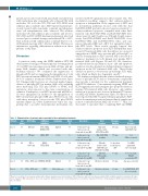
Table 2. Characteristics of patients who responded to the combination treatment.
Patients
Case 1
Case 2
Case 3
Case 4
Case 5
Case 6
Age FLT3 Status (years)
81 ITD mutb
63 ITD mut
24 ITD + D835 mut
38 ITD mut
Additional mutationsa
TP53, IDH2, RUNX1, WT1
HRAS. WT1
NRAS, TET2, IDH1
RUNX1
Karyotypes Blasts (%)
Diploid 67
t(6;9) 10
Diploid 68
Miscellaneous 80
Diploid 66
Miscellaneous 94
Dose level
-Selinexor 80 mg twice per week -Sorafenib 400 mg BID
-Selinexor 60 mg twice per week -Sorafenib 400 mg BID
-Selinexor 80 mg twice per week -Sorafenib 400 mg BID
-Selinexor 60 mg twice per week -Sorafenib 400 mg BID
-Selinexor 60 mg twice per week -Sorafenib 400 mg BID
-Selinexor 60 mg twice per week -Sorafenib 400 mg BID
Best response
CRp with undetectable FLT3-ITD by PCR
CRp, FLT3 PCR pending
CRi with undetectable FLT3-ITD and FLT3-D835 by PCR
CRi with FLT3 positive by PCR
Hi-blast reduction
Hi-blast reduction
Response duration
180 days
75 days
60 days then to autologous SCT
60 days
35 days
35 days
Prior therapies
Aza+sorafenib, Anti-CD25 trial
7+3, CLIA+sorafenib
7+3, MEC,
quizartinib, IA+crenolanib
7+3+sorafenib, GCLAC +sorafenib, SCT
Aza, aza+sorafenib
7+3, MEC, MUD, SCT, Dac+sorafenib,
78 ITD+D835mut DNMT3A,RUNX1
50 ITD + D835 mut WT1
aBased on screening of bone marrow samples with a 28-gene mutation panel. bmut: mutations. PCR: polymerase chain reaction; Aza: azacytidine; CLIA: cladribine idarubicin, cytarabine; IA: idaru- bicin,cytarabine; MEC:mitoxantrone,etoposide,cytarabine;GCLAC:granulocytecolony-stimulatingfactor,cladribine,cytarabine;SCT:stemcelltransplantation;MUD:matchedunrelateddonor; Dac: decitabine.
1650
haematologica | 2018; 103(10)
