
Page 74 - 2018_10-Haematologica-web
P. 74
E. Gars et al.
olds that can be used to accurately diagnose most cases of HLH: one granulocyte or two nucleated erythroid cells per 1000 nucleated cells. In addition, we utilized classification and regression tree analysis to identify the best combina- tion of variables to create an even more specific and pre- dictive model for discriminating patients with HLH. Finally, we showed that the presence of multiple nucleat- ed cells within a single hemophagocyte was additionally predictive of a diagnosis of HLH among patients present- ing with clinical findings of concern for the disease.
The initial description of virus-associated hemophago- cytic syndrome (VAHS) described florid hemophagocy- tosis in 19 cases of immunosuppressed and previously healthy patients presenting with clinical features com- patible with HLH.11 In 60 consecutive bone marrow aspirates used for comparison, they found erythrophago- cytosis in 29 of 60 cases; however, the degree of phago- cytosis was “never of a degree to be confused with the VAHS”. This study conducted prior to the development of the HLH-2004 criteria provided initial evidence of the association of hemophagocytosis with VAHS (what we
would now call HLH), and showed that the finding of erythrophagocytosis is frequently identified in bone mar- row aspirates of patients without HLH, demonstrating a lack of specificity in isolation of clinical findings.
We conducted a similar non-blinded evaluation of 87 bone marrow aspirates from patients with de novo and post-treatment myeloid and lymphoid malignancies, benign cytopenias, and negative staging marrows, and identified predominantly erythrophagocytosis of mature RBCs in 39% of these cases. Applying the quantitative threshold determined from our analysis for engulfed non-nucleated RBCs (erythrophagocytes) to this dataset, we find only 3 of the 87 patients (5%) would satisfy this morphological criterion (4 per 1000 cells). Similarly, if we apply the quantitative threshold for granulocytes (1 per 1000 cells), only 4 of the 87 patients (4%) would satisfy this morphological criterion. Therefore, although we can indeed find rare examples of erythrophagocytosis in aspirate smears of a large minority of cases, the applica- tion of quantitative thresholds reveals a low incidence of clinically significant hemophagocytosis in a patient pop-
A
B
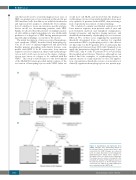
Figure 4. Classification and regression trees (CART). Lineages were dichotomized based on the threshold values obtained from Table 3 and entered into the CART. Lineages are ordered in terms of relative importance to hemophagocytic lymphohistiocytosis (HLH), where variables on the higher levels are deemed more important. The shaded area in each box corresponds to the probability of having an HLH diagnosis based on the path that leads to it. All four lineages were entered into the CART in (A), while lymphocytes were excluded from the CART in (B).
1640
haematologica | 2018; 103(10)
