
Page 70 - 2018_10-Haematologica-web
P. 70
E. Gars et al.
ABCD
EFGH
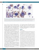
Figure 1. Examples of hemophagocytosis in patients with hemophagocytic lymphohistiocytosis (HLH). (A) Histiocytes in patients with HLH often display rounded contour with cytoplasmic projections. (B-D) Hemophagocytes with a single ingested mature red blood cell (RBC), nucleated RBC progenitor, and granulocyte, respec- tively. Hematopoietic progenitor cells (HPCs) often contain single nucleated hematopoietic cells in addition to multiple mature RBCs (E); however, the presence of multiple nucleated cells within the cytoplasm of a single HPC (F and G) is highly predictive of the diagnosis of HLH. (H) An example of a histiocyte with degenerating nuclear debris, indistinct cytoplasmic contour, and equivocal intracytoplasmic nucleated RBCs that we do not consider to be a definite hemophagocyte.
tion tests.1 The most widely used diagnostic criteria for HLH were developed for inclusion in the HLH-2004 trial which requires genetic evidence of a mutation associated with HLH or fulfillment of 5 of 8 clinical criteria including fever, splenomegaly, bicytopenia, hypertriglyceridemia or hypofibrinogenemia, evidence of hemophagocytosis in bone marrow or other tissues, low or absent NK-cell activ- ity, elevated ferritin, and elevated soluble IL-2 receptor.3 Although not validated for adults, these HLH-2004 criteria are broadly applied in patients of all ages.
Pathologists play a critical role in the diagnostic workup of patients suspected of having HLH. Bone marrow exam- ination is performed to evaluate for hemophagocytosis, identify underlying malignancy, and exclude benign or neoplastic mimics. The presence of hemophagocytosis in the marrow fulfills one of the HLH-2004 diagnostic crite- ria; however, no accepted diagnostic threshold or report- ing guidelines have been established. The lack of evi- dence-based guidelines leads to considerable uncertainty among pathologists as to what degree of hemophagocyto- sis is sufficient to satisfy this criterion. Adding to the chal- lenge is that hemophagocytosis is not specific to the diag- nosis of HLH in the absence of other clinical features of the disease. Rare erythrophagocytosis is commonly seen in bone marrow aspirates and increased hemophagocyto- sis may be encountered in the setting of sepsis, blood transfusions, hematopoietic transplantation, chemothera- py, and myelodysplastic syndrome.8-11
Given the lack of a defined threshold to fulfill the crite- rion for diagnosis of HLH, we designed this retrospective study to interrogate whether quantitative or qualitative morphological features of hemophagocytosis in bone mar- row aspirates are predictive of the eventual diagnosis of HLH. We identified a cohort of patients presenting with clinical characteristics that were of concern for HLH and their aspirates were examined blindly.
Hemophagocytes were enumerated per 1000 nucleated
cells according to the lineage of their ingested hematopoi- etic contents [mature red blood cells (RBCs), nucleated RBCs (nRBCs), granulocytes, and lymphocytes] (Figure 1). In addition to quantitative features, we evaluated a binary morphological feature, the presence of multiple nucleated cells within a single hemophagocyte, as a possible predic- tive characteristic of HLH.
Methods
Patient selection
We searched the pathology Laboratory Information Service data- base (Powerpath) using the following keywords: “hemophagocytic lymphohistiocytosis”, “hemophagocytosis”, “erythrophagocyto- sis”, and “HLH”. This search returned 258 results between the dates 1st January 2013 and 7th January 2017, and included text from any- where within the diagnostic report including the provided clinical information, microscopic description, diagnostic line, and/or diag- nostic comment (Figure 2). These patients’ medical records were reviewed by EG to assess whether clinical suspicion for HLH was present at the time of bone marrow aspiration, either indicated on the specimen requisition form (i.e. “rule out HLH” or “concern for HLH”) or listed in the differential diagnosis in the electronic medical record (EMR) within one week prior to biopsy. Demographic infor- mation, clinical characteristics, diagnostic impressions, pathological features, and laboratory values at the time of biopsy were collected for each patient. Patients were classified as “HLH” and “non-HLH” based on the diagnostic impression of the consulting hematologists described in the clinical notes. The final diagnosis in all cases was determined based on the HLH-2004 criteria in conjunction with the overall clinical picture. Patients were excluded from the analysis if hemophagocytosis was incidentally noted independent of clinical concern for HLH, slides were not available for review, HLH was considered but the diagnosis was equivocal after workup, or a doc- umented history of HLH-directed treatment was noted prior to biopsy. This study was approved by Stanford University’s institu- tional review board.
1636
haematologica | 2018; 103(10)
