
Page 160 - 2018_10-Haematologica-web
P. 160
B.A. Williams et al.
A
B
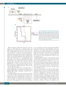
Figure 5. iNK-92 therapy of primary acute myeloid leukemia (AML) xenografted NOD/SCID gammanull (NSG) mice. 3x10⁶ pri- mary AML cells were injected intravenously (i.v.) via tail vein into irradiated NGS to establish disease in control (n=5) and therapy (n=5) mice. iNK-92 given intraperitoneally (i.p.) 20x206 weekly for six weeks were used to treat AML xenografted mice starting ten days after inoculation (A). Mice were monitored for signs of leukemia and sacrificed at humane end points (B). Kaplan- Meier survival curves were generated to compare survival in control and treatment groups (P=0.0566).
Only two studies to date have looked at the in vitro sen- sitivity of CD34+CD38– LSCs to immune effector cell killing. In the first, lymphokine-activated killer (LAK) cells and allogeneic lymphocytes exerted a modest cytotoxic effect on AML LSCs comparable to the effect on the non- stem cell fraction.23 In a more recent study, endogenous single killer immunoglobulin-like receptor (KIR)-express- ing NK cells, mismatched for the HLA of primary AML targets, showed equivalent killing of LSCs and blasts with either the chromium release or methylcellulose-based cytotoxicity assays.24 Here, we demonstrate preferential killing of LSCs versus bulk leukemia by NK-92, not shown by these other studies using a more rigorously controlled methylcellulose cytotoxicity assay than that used by Lankencamp et al. This is consistent with our work on NK-92 treatment of multiple myeloma (MM) cell lines showing that NK-92 preferentially kills clonogenic MM cells over bulk tumor cells.25
To pursue more in-depth studies of the effectiveness of NK-92 in killing LSCs, we developed an animal model of primary human AML by using NSG mice infused with a primary AML sample containing a small fraction of CD34+CD38– cells.
Secondary transplantation is the current gold standard to determine the effect of small molecules on LSCs26 but is rarely used to evaluate cellular therapies for leukemia. We attempted this assay by transplanting bone marrow from control and NK-92 treated mice into new recipients to assess the impact on individual mice rather than on pooled cells. BM engraftment occurred in all AML-only
cohort secondary mice, while one mouse from the iNK-92 group was leukemia free, with engraftment at the back- ground levels of non-injected mice. While the average BM engraftment of secondary transplant mice in the therapy groups was less than the control, this was not statistically significant. However, the LSC fraction was significantly decreased in secondary recipients, providing some evi- dence of NK-92 cytotoxicity against LSCs in the second- ary transplant assay.
AML-xenografted NSG mice were effectively treated with NK-92 infusions, leading to improvement in survival versus controls, confirming previous work.11 We accom- plished this with lower doses of NK-92 on a less com- pressed schedule than the original study, and without the use of IL-2 in the regimen. Irradiated NK-92 could prolong survival in mice, but was less effective than the non-irra- diated cells. We postulate that the reason for this reduc- tion in therapeutic efficacy is the lack of ability for the cells to proliferate in vivo.
We have demonstrated for the first time that irradiated NK-92 improves survival in an AML xenograft model, which has translational relevance given that only irradiat- ed NK-92 is administered to patients in phase I trials. However, recognizing that the effect of iNK-92 on improving survival in vivo is modest, we wished to add the mechanism of ADCC to cell killing by using the genetical- ly modified CD16+NK-92 in combination with a mono- clonal antibody. CD16+NK-92 has been combined with rituximab to enhance killing of CD20+ malignant cells, showing its potential to enhance the killing of cells
1726
haematologica | 2018; 103(10)
