
Page 160 - 2018_09-Mondo
P. 160
C.C.F.M.J. Baaten et al.
analysis of platelet responsiveness was performed during the decreasing period of platelet count (50-11 x109/L and ≤10 x109/L), and the recovery of platelet count (11-50 x109/L). The latter was defined as a sustained increase in the platelet count (observed for patient care), independent of platelet transfusion. Of the eight patients included in this category, three had received an autologous transplant and one patient an allogeneic stem cell transplant, prior to recovery. In the decreasing period, integrin activation and P-selectin expression following stimulation with thrombin or CRP-XL were comparable in patients with platelet counts in the range of 50-11 x109/L and ≤10 x109/L (Figure 2). In contrast, platelet responsiveness to thrombin and CRP-XL significantly improved in the case of count recov- ery (P<0.001). For stimulation with ADP, these differences were less pronounced, with only P-selectin expression increased during count recovery. These results indicated that platelet count alone is not a good marker of platelet activity.
For five patients (one AML, three multiple myeloma, one lymphoma), blood samples could also be analyzed at an earlier time point, i.e., after the stop of chemotherapy, but before severe thrombocytopenia occurred. Remarkably, in all these samples, platelet function was within the normal range for the three agonists (integrin activation 69-86%, P-selectin expression 49-85%). Furthermore, in vitro treatment of control blood with clin- ically relevant concentrations of cytarabine and/or mel- phalan did not affect platelet reactivity (Online Supplementary Figure S3A,B). These results argue against a direct effect of chemotherapeutics on the platelet activa- tion properties.
Impaired platelet spreading and Ca2+ signaling of platelets after chemotherapy treatment
To further characterize the patient platelets, they were allowed to adhere and spread for ten minutes on a fibrino- gen surface, interacting with platelet integrin aIIbβ3. The
observed morphology of the cells was divided into three stages: 1) formation of filopodia, 2) formation of lamel- lipodia, and 3) full spreading. Most of the platelets from control subjects were in stages 2-3, while the patient platelets predominantly stayed in stage 1 (forming filopo- dia only), with few platelets being fully spread (Figure 3A). The patients’ platelets displayed a slightly decreased expression of glycoprotein (GP)Iba and GPVI, but not in integrin β3 expression (data not shown). This suggested a diminished integrin activity and outside-in signaling in the patient platelets.
We further examined agonist-induced Ca2+ signaling after loading the platelets with Fluo-4. Stimulation with thrombin or CRP-XL induced only a small rise in [Ca2+]i in patient platelets when compared to control platelets (Figure 3B,C). On the other hand, the [Ca2+]i rise induced by thapsigargin (an inhibitor of endoplasmic reticulum Ca2+-ATPases), as a measure of Ca2+ store content,21 was similar for patient and control platelets. Together, this pointed to a defective agonist-induced Ca2+ signaling machinery, independently of receptor type (i.e., PAR1/4 or GPVI receptors).
Impaired mitochondrial bioenergetics but no apoptosis in platelets after chemotherapy
Given the cytotoxicity of chemotherapeutic com- pounds, we evaluated if patient platelets showed charac- teristics of apoptosis, since this process is known to lead to dysfunctional signaling.22 As a marker of apoptosis, PS exposure was determined by fluorescein isothiocyanate (FITC)-annexin A5 binding. In contrast to control platelets, the patient platelets were prone to expose PS upon short- term storage without external stimuli (Figure 4A). Upon stimulation with the BH3 mimetic ABT-737, triggering the intrinsic pathway of apoptosis,22 PS exposure was initially accelerated in the patient platelets, when compared to control platelets (Figure 4B). As expected, preincubation with the pan caspase inhibitor quinoline-val-asp-difluo-
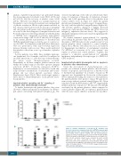
A
B
Figure 1. Variable impairment of integrin aIIbβ3 activation and P-selectin expression in stimulated platelets from cancer patients with thrombocytopenia after chemotherapy. Washed platelets (10x109/L) from healthy control subjects (healthy ctrl) and thrombocytopenic patients receiving chemotherapy were activated with thrombin (4 nM), CRP-XL (10 μg/mL) or 2MeS-ADP (1 μM) in the presence of 2 mM CaCl2. After 15 min activation, integrin aIIbβ3 activation (A) and P-selectin expression (B) were measured by flow cytometry using PAC-1 and anti-P-selectin antibody, respectively. Medians with IQR; data from 52 patients (25 AML/ALL, 12 multiple myeloma, 13 lymphoma, two other), 27 healthy controls, ***P<0.001. CRP: collagen-related peptide; ADP: adenosine diphosphate.
1560
haematologica | 2018; 103(9)
