
Page 57 - Haematologica August 2018
P. 57
Activity of SL-401 in AML and MDS
AC
B
D
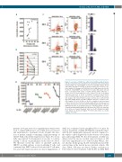
Figure 3. Low levels of CD123 are sufficient for SL-401-mediated cytotox- icity. (A) Correlation between CD123 levels and SL-401 induced cytotoxicity in patient AML cells. CD123-MESF on vehicle-treated AML samples (N=13) and viability after 48 hr of SL-401 (1 μg/ml) treatment were used for corre- lation analysis. (B) Changes in CD123 molecules in AML blasts after SL-401 treatment. CD123-MESF was determined on viable AML blasts after 24 hours of treatment with vehicle or SL-401 (N=13). Mean difference = 11086 (95% CI for mean difference: 2006, 20166), P=0.021. (C-D) CD123 expression and effect of SL-401 on umbilical cord blood derived CD34+ cells. (C) CD34 positive selected cells were used for multicolor flow cytom- etry analysis and colony formation assays. Colonies were counted 10-14 days after plating CD34+ cells with continuous presence of vehicle or SL- 401(1 μg/ml) P=0.019. (D) Effect of SL-401 on umbilical cord blood liquid cultures. Non-enriched Cord blood samples (N=4 CB) were ficoll processed to obtain mononuclear cells and cultured in RPMI media with 20% FBS and GM-CSF, SCF and IL-3 (10 ng/ml) in presence of SL-401 and the cells were counted and immunophenotyped after 48 hours. Live CD34+ cell counts per ml of culture are shown in the graph. Vehicle vs. SL-401 10ng/ml not signif- icant; Vehicle vs. SL-401 100ng/ml; P=0.0356 and Vehicle vs. SL-401 1 μg/ml; P=0.0089.
patients (n=3) pre-tested for engraftment potential were used to engraft NRGS mice (one AML donor per mouse) and randomized to treatment cohorts. Despite the selec- tion of patient samples containing a high percentage of CD123+AML cells (Online Supplementary Figure S7), we ini- tially encountered T-cell expansion in vivo due to contam- inating T cells in our preliminary studies (data not shown). We therefore tested the effect of anti-CD3 antibody (OKT3) on AML cultures in vitro in ablating T cells, and confirmed that OKT3 reduced both absolute T-cell num- bers and CD3 expression (data not shown). Engraftment of
AML was confirmed in the peripheral blood of mice by week 4. Treatment of (AML 28/AML29) xenografted mice with SL-401 significantly increased survival compared to the vehicle control (Figure 5A). In the absence of busulfan preconditioning (AML 30), the development of AML was delayed and the mouse reached removal criteria at 102 days after engraftment (versus with busulfan 48days; data not shown). In this group, SL-401 treatment increased the survival time in the treated mouse (survival: vehicle, 102 days; SL401, 154 days; not shown). Further, evaluation of bone marrow after treatment with SL-401 in AML PDX
haematologica | 2018; 103(8)
1293
