
Page 54 - Haematologica August 2018
P. 54
R. Mani et al.
Coulter) after establishing calibration curve for each experiment per the manufacturer’s instructions. These CD123-MESF experi- ments were performed at the OSU Wexner Medical Center Clinical Cytometry Facility. Flow cytometric data were analyzed using Kaluza software (Beckman-Coulter).
AML Patient-Derived Xenograft (PDX) models
Animal experiments were performed under a protocol approved by the OSU Institutional Animal Care and Use Committee (IACUC) in NRG-SGM3 (NRGS) mice. Detailed in Online Supplementary Methods.
Statistics
Detailed in Online Supplementary Methods. Results
AML cells express varying levels of CD123 and are sensitive to SL-401
While more than 80% of AML cases show CD123 expression on blasts,28 the levels of expression are variable. Therefore, we tested the cell surface expression of CD123 on primary AML blasts. As expected, CD123 was found on the majority of AML samples tested (Table 1, and Online Supplementary Figure S1). Rarely, CD33-/CD123+ AML cell populations were also observed (Online Supplementary Figure S2A). SL-401 induced potent cytotox- icity on AML primary cells as seen by dose-dependent reduction in viability of AML cells (Figure 1A) and absolute cell numbers (Online Supplementary Figure S2B). The clonogenicity of AML samples was also significantly reduced by SL-401 as seen in colony forming assays (Figure 1B). We next determined if the activity of SL-401 was diminished in the presence of IL-3. In AML cell cul- tures containing 10ng/ml IL-3, a concentration > 100 fold of physiological levels, SL-401 was effective in inducing cytotoxicity and reducing the cell counts (Online Supplementary Figure S2 B-C). However, SL-401 (100ng/ml) cytotoxicity was inhibited by IL-3 in dose dependent manner, especially at doses >1000 fold of physiological levels, proving target specificity (Online Supplementary Figure S2D). Blasts derived from high-risk FLT3-ITD mutated AML often express high levels of CD123,28 so we next tested the activity of SL-401 on AML primary cells and cell lines with this mutation. Importantly, high risk FLT3-ITD+ AML tend to express similar or higher than median CD123-MESF (22825) and responded to SL-401 (Figure 1C). The FLT3-ITD+AML cell lines MV4-11 and MOLM-13 express CD123 with other myeloid markers CD45, CD33 (Figure 1D) and SL-401 strongly inhibited the growth of these cell lines (P<0.0001 for dose trends of MOLM-13 and MV4-11) (Figure 1E). However, SL-401 did not induce cytotoxicity in CD123 cell line K562 proving target specificity (Online Supplementary Figure S3A). Interestingly, cell density did not affect cytotoxicity in CD123+ MV4-11 cells (Online Supplementary Figure S3B).
SL-401 overcomes autologous stromal cell protection in co-cultures
As AML cells and their progenitors derive growth and survival support from the bone marrow stromal cells and microenvironment,22,23 we evaluated the effect of stromal protection on SL-401-induced cytotoxicity. Although
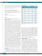
Table 1. Acute myeloid leukemia blasts express varying levels of cell surface CD123.
AML
AML 1
AML 2 AML 3 AML 4 AML 5 AML 6 AML 7 AML 8 AML 9 AML 10 AML 11 AML 12 AML 13 AML 14 AML 15
AML 16
Blasts (B)
2.03
1.73
2.45
2.35
1.48
1.29
1.77
1.59
1.13
1.02
0.83
1.44
1.60
1.29
0.46
0.41
CD123-MFI
Lymphs (L) B/L Ratio
0.28 7.38
0.32 5.49
0.58 4.21
0.61 3.86
0.58 2.53
0.28 4.56
0.35 5.00
0.40 3.94
0.37 3.10
0.36 2.81
0.40 2.06
0.37 3.86
0.37 4.32
0.39 3.33
0.35 1.30
0.37 1.11
CD123-MESF on blasts
ND
ND
13672
10692
4396
ND
47791
61863
66972
33137
14117
22825
19297
63204
35980
18636
1290
CD123-MFI and CD123-MESF on blasts and lymphocytes (N=16) are shown. MFI: Mean Fluorescence Intensity; MESF: Molecules of Equivalent Soluble Fluorochrome; ND: Not determined.
MV4-11 cells did not receive additional protection, the protective effect of stromal HS-5 on MOLM-13 and AML cells was reduced by SL-401 treatment (Figure 2 A-B). We next tested the effect of SL-401 on AML cells cultured on primary autologous bone marrow stromal cells from AML patients. For this purpose, we derived MSC from individ- ual AML patient marrow samples. MSCs grown in flasks exhibited stellate- to spindle-shaped large cells and were adherent. Purity and phenotype were confirmed by multi- color flow cytometry (Online Supplementary Figure S4A). Importantly, SL-401 was able to reduce the growth of AML regardless of culture with autologous MSC (Figure 2C and Online Supplementary Figure S4B).
Low levels of CD123 are sufficient for SL-401 mediated cytotoxicity
Although earlier studies utilizing variant/wild type IL-3- diphtheria toxin have reported direct correlation of cyto- toxicity to the levels of IL-3R subunits, the clinical trials have shown intriguing results with lack of correlation between pharmacokinetics and pharmacodynamics.19, 20,29- 34 To study this relationship between SL-401 and its target, we compared the sensitivity of AML cells to SL-401 vs. their expression of CD123. As shown in Figure 3A, there was no significant correlation between the relative viabil- ity of AML samples treated with SL-401 for 48 hours with their CD123 expression (CD123-MESF on AML cells). Interestingly, AML cells expressing as few as 10,000 CD123 molecules were still sensitive to SL-401-mediated cytotoxicity. As leukemic cells can acquire resistance through target down regulation during the course of treat- ment, we sought to determine if SL-401 modulated cell surface CD123 levels after exposure. Although SL-401 treated samples had slightly reduced CD123 (CD123
haematologica | 2018; 103(8)
