
Page 22 - Haematologica August 2018
P. 22
1258
M. de Charette et al.
Genetic alterations
Direct, genetic alterations leading to MHC-II loss are mostly seen in DLBCL of immune-privileged sites. PTL and PCNSL have lost HLA-DR in 61% and 46% of cases, respectively.48 In contrast with other types of DLBCL, genetic lesions of MHC-II genes represent the main mech- anism of HLA-DR loss:48,49 MHC-II is mutated in 78% of PTL and 50% of PCNSL.49 Transcription factors seem to be rarely implicated in HLA-II loss in PTL: CIITA and FOXP1 rearrangements are present in only 10% and 7% of cases, respectively.50
It is noteworthy that, when expressed, MHC-II may drive inhibitory signals. Indeed, lymphocyte-activation gene 3 (LAG-3), a member of immunoglobulin superfami- ly expressed on tumor infiltrating lymphocytes (TILs),51 binds to MHC-II with greater affinity than CD4, leading
to the inhibition of TCR signaling, proliferation and cytokine secretion by antigen-specific T cells. Exhausted LAG-3 positive TILs are present in the immune infiltrate of FL, DLBCL and HL (mostly in EBV positive cases, mixed cellularity and rich lymphocyte subtypes).52,53 Furthermore, circulating CD4 T cells from HL patients with active disease express LAG-3 at higher levels than healthy donors or patients in long-term remission.53
Antigen processing machinery alterations
GILT and HLA-DM are enzymes of the antigen process- ing machinery (APM), located in the endocytic compart- ment of APC and B cells. Both are down-regulated by cMYC, leading to a defective antigen presentation that can be restored in vitro by cMYC inhibitors.54
GILT generates epitopes to be loaded on MHC-II. In
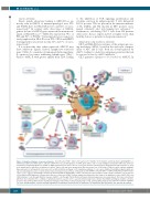
Figure 1. Lymphoma immune evasion mechanisms. (Top left panel) "Hide". Tumor cells may become “invisible” to the immune system by down-regulating MHC, co- stimulatory (CD80 and CD86) and/or adhesion (CD54) molecules. Downregulation of CD58 allows tumor cells to escape killing by natural killer (NK) cells, which are activated by self-missing signal (loss of MHC-I). (Right panel) "Defend". Tumor cells are seen by the immune system but avoid destruction through resistance to apop- tosis signals and/or expression of inhibitory receptors. Tumor cells may resist apoptosis by different means: loss of FAS and/or TRAIL receptors (extrinsic pathway), hyperexpression of anti-apoptotic molecules such as BCL-2 (intrinsic pathway) or PI9 (Granzyme pathway). T cells can be inhibited by inhibitory ligands which are expressed by lymphoma cells or cells from their microenvironment such as PD-L1 or PD-L2/PD-1, LAG-3/MHC-II, CTLA-4/CD80 or CD86 and HLA-G/ILT. CD47 sends a “don’t eat me” signal to macrophages and DCs by interacting with its ligand SIRPa. Tumor cells may also express FAS-L to induce death of immune cells. Some mol- ecules expressed by lymphoma cells may have dual roles: expression of MHC-II allows antigen presentation but also binds to the inhibitory receptor LAG-3; CD80 and CD86 stimulate T cells through CD28 but may also inhibit T cells through CTLA-4. (Bottom left panel) Immunosuppressive microenvironment. The tumor cells interact with their microenvironment to contribute to lymphoma immune evasion. IL-10 is a potent immunosuppressive cytokine that inhibits priming by dendritic cells (DC), promotes Th2 and Treg differentiation and M2 macrophages; TGF-b induces exhausted phenotype of CTL and Treg differentiation; IDO suppresses cytotoxic T lympho- cyte (CTL) and NK immune response through degradation of tryptophan and production of kynurenine. Trp: tryptophan; Kyn: kynurenine; Gal: galectin; Ag: antigen.
haematologica | 2018; 103(8)
