
Page 174 - Haematologica August 2018
P. 174
M.T. Georgescu et al.
altered expression due to FVIII+Dex treatment (Figure 7A, Online Supplementary Table S1 and Table S2). There were no differences between the splenic mRNA transcription profiles of FVIII+Dex and FVIII mice (Figure 7B). There were also no differences between the thymic and splenic mRNA transcription profiles of FVIII and HBSS mice (Online Supplementary Figure S2).
Discussion
We sought to determine whether Dex, when adminis- tered during initial FVIII exposure, could promote immunologic tolerance to FVIII in HA mice. Our experi- ments indicate that both E17KO/hMHC and E16KO FVIII+Dex mice were less likely to develop anti-FVIII IgG than FVIII mice after initial exposure to FVIII. Although E17KO/hMHC mice can have inherent tolerance to FVIII, the ability of Dex to also reduce inhibitor development in E16KO mice is encouraging. While any immediate effect might have been due to transient immunosuppression, the reduced incidence of anti-FVIII IgG in E17KO/hMHC FVIII+Dex/FVIII mice after re-exposure to FVIII at six, and especially 16, weeks suggests that long-lasting tolerance to FVIII can be promoted. Furthermore, this tolerance is spe- cific to FVIII since these mice mount a robust response to a structurally unrelated antigen (human VWF) despite remote exposure to Dex.
Especially noteworthy is the reduced anti-FVIII immune response after Dex exposure in mice whose re-exposure to FVIII was accompanied by LPS. LPS administration with FVIII has been reported to yield anti-FVIII IgG in 100% of E17KO/hMHC mice,14 an effect also observed in our experiments. Compared to FVIII/FVIII+LPS mice, FVIII+Dex/FVIII+LPS mice demonstrated a markedly reduced anti-FVIII immune response, although this effect did not reach statistical significance due to small numbers.
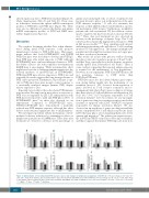
We investigated potential cellular mechanisms of Dex- mediated tolerance induction by examining its effect on lymphocyte populations of E16KO mice. Three days after treatment we observed a decrease in the percentage of
splenic and circulating B cells. A subset of splenic B cells has been shown to play a role in the initiation of the anti- FVIII immune response.19 B cells also maintain this response as their inhibition has been identified as a poten- tial mechanism of ITI in mice.20 Furthermore, in inhibitor patients who fail conventional ITI, the addition of ritux- imab to target B cells has been shown to increase ITI effi- cacy.21 Three days post-treatment we also observed an increase in the percentage of thymic Tregs. This T-cell subset has been repeatedly implicated in tolerance to FVIII in HA mouse models.18,22,23 Tregs simultaneously interact with antigen-presenting cells and effector T cells, resulting in effector T-cell suppression. The changes in lymphocyte populations following Dex treatment were no longer pres- ent three weeks post-treatment.
Our results are in line with previous studies showing that glucocorticoids can induce apoptosis of B and T cells13 and that Tregs, especially those in the thymus, are prefer- entially spared from Dex-induced cell death.24 There is some evidence suggesting that repeated antigen exposure is required for the maintenance of Treg populations.25 However, in our experiment, FVIII+Dex/intFVIII mice did not maintain tolerance to FVIII better than FVIII+Dex/FVIII mice.
FVIII+Dex mice also had altered thymic gene expres- sion, giving further insight into the mechanism of our treatment protocol. We observed down-regulation of genes involved in T-cell receptor formation and re- arrangement (Cd4, Rag1, Rag2), genes coding for cytokines that drive effector T-cell proliferation and maturation (Il9, Il12b, Il13,26 Il16,27 Il2728) and genes responsible for T-cell activation (Cd40lg,29 Lck30). In contrast, we saw up-regula- tion of genes encoding for scavenger receptors involved in clearance of apoptotic cells (Cd36,31 MARCO32) and genes responsible for thymic involution (Pparg33). We also observed an up-regulation of genes encoding extracellular matrix components (Fn134) and adhesion molecules (Ita2b, Cdh5) that may play a role in regulating thymocyte devel- opment and migration.35 The splenic gene expression pro- file in FVIII and HBSS control mice were almost identical, and thus it appears that intense FVIII exposure alone does
ABC
DEF
Figure 6. Administration of Dex during initial FVIII exposure causes early changes in lymphocytes populations of E16KO mice. The percentage of A. B cells, B. T cells and C. Tregs in the thymus, spleen and blood three days after treatment with HBSS, Dex, FVIII or FVIII+Dex. The percentage of D. B cells, E. T cells and F. Tregs in the thymus, spleen and blood three weeks after treatment with HBSS, Dex, FVIII or FVIII+Dex. n=3-7 for each condition. *P<0.05, **P<0.01, ***P<0.001.
: HBSS; : Dex; : FVIII; : FVIII+Dex.
1410
haematologica | 2018; 103(8)
