
Page 156 - Haematologica August 2018
P. 156
1392
A. Roberto et al.
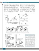
low levels of cCD56dim NK cells early after hHSCT were counterbalanced by a significant expansion the uCD56dim NK cell subset which, instead, was present at low levels in PB under homeostatic conditions. uCD56dim NK cells start- ed to statistically increase in the recipients compared to their related donors in the second week after the trans- plant, outnumbered all other NK cell subsets in the second and third week, and returned to normal level only eight weeks after hHSCT. Different from Human Leukocyte Antigen (HLA)-matched HSCT,26 CD56bright/CD16pos NK cells did not increase in frequency at any time-point in
hHSCT recipients compared to their related donors (Figure 1C,D). We and others have reported that the infec- tion/re-activation with human cytomegalovirus (HCMV) greatly influences the homeostasis and ontogenesis of NK cell subset and induces the expansion of CD56neg/CD16pos NK cells. This phenomenon is particularly relevant in the immune-compromised recipients receiving allogeneic HSCT.14 Although 23 of the 30 (77%) transplanted patients recruited for this study experienced a HCMV infection/reactivation starting from day 29 post hHSCT up to day 64 following the transplant (Online
A
B
C
D
Figure 1. Kinetic of NK cell subset immune reconsti- tution after haploidentical HSCT. (A) Summary graph showing the absolute counts (cells/μL) of circulating natural killer (NK) cells (mean ± SEM) from hematopoietic stem cell healthy donors (HDs) and their related recipients at different time-points after haploidentical HSCT (hHSCT). (B) Representative example of flow cytometry dot plots showing the com- plete chimerism of HD-derived HLA-A2neg NK cells reconstituting an HLA-A2pos recipient (upper line) after four and eight weeks from hHSCT (lower line). (C) Representative example of flow cytometry dot plots showing the kinetic of HD-derived NK cell subset dis- tribution in the recipient after two, three, four and five weeks from hHSCT. (D) Summary statistical graph showing the frequency (median ± SEM) of convention- al CD56bright/CD16neg-low (cCD56bright), CD56bright/CD16pos, conventional CD56dim/CD16pos (cCD56dim) and uncon- ventional CD56dim/CD16neg (uCD56dim) NK cell subsets in the peripheral blood (PB) and bone marrow (BM) of 30 hematopoietic stem cell HDs compared to their counterparts in the blood of the related recipients up to six months after hHSCT. *P<0.05.
haematologica | 2018; 103(8)
