
Page 65 - Haematologica Vol. 109 - July 2024
P. 65
ARTICLE - Leukemia-induced interferon signature in stroma M.W.E. Smeets et al.
exposure to ETV6-RUNX1-positive cells did not markedly decrease the viability of primary ETV6-RUNX1 (nor B-other) BCP-ALL cells in 3-day culture assays (Figure 5). Moreover, silencing the key regulator of the IFN-signaling pathway, i.e., STAT1, in MSC also did not result in altered ALL via-
A
bility after 40 or 120 hours of co-culture, while there was a reduction of ≥60% in STAT1 protein expression (Online Supplementary Figure S12). Thus, the viability of leukemic cells was not affected by interference with IFN signaling via silencing of individual IFN-related genes.
B
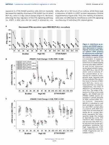
Figure 6. Interferon-α se- cretion and IFNAR1 expres- sion decreased in mesen- chymal stromal cells after co-culture with primary B-cell precursor acute lym- phoblastic leukemia cells. (A) Interferon-alpha (IFNα) concentration in superna- tants collected after 40 hours from 15 mesenchymal stromal cells (MSC) (MSC#1- 3) and B-cell precursor acute lymphoblastic leuke- mia (BCP-ALL) (ALL#1-15) mono- and co-cultures. IF- Nα concentration is cor- rected for medium back- ground level. Squares and circles indicate IFNα secre- tion level in the sum of MSC and BCP-ALL mono-cul- tures and their co-cultures, respectively. The limit of detection is indicated by a solid line. The range of de- tection for IFNα is 0.5-2,503 pg/mL. (B) IFNAR1 and (C) IFNAR2 mRNA levels ex- pressed in paired samples of MSC sorted after mo- no-culture (squares; MSC#1-3 indicated in red, blue, and green, respective- ly.) and MSC sorted after co-culture with BCP-ALL cells from 15 individual pa- tients (#1-15; circles). Box- plots represent the inter- quartile range, the median is depicted by a line. IFN: interferon; MSC: mesenchy- mal stromal cells; BCP-ALL: B-cell precursor acute lym- phoblastic leukemia; HD: high hyperdiploid; FDR: P-value false discovery rate; FPKM: fragments per ki- lobase million.
C
Haematologica | 109 July 2024
2079
