
Page 355 - Haematologica Vol. 109 - July 2024
P. 355
CASE REPORT
The initial increase in size, erythema around these skin lesions and worsening pain followed by resolution of these lesions was concluded to be an example of TFR second- ary to talquetamab. The patient was only on prophylactic antimicrobials and there was no discharge from these fun- gating lesions and clinically we did not suspect cellulitis. The patient’s lesions were consistent with prior biopsy proven EMD. Most significantly there was a clear tempo- ral relationship to treatment, and a correspondence with serological myeloma markers. The rise in LDH (a frequent surrogate for disease activity and tumor lysis) and even a slight rise in the κ light chains corresponded with the ex- pansion of skin lesions. The skin lesions resolved completely as the light chain ratio normalized, LDH fell and patient achieved a biochemical CR. The CR in this penta-refrac- tory, unfavorable cytogenetics patient continues to affirm the effectiveness of novel BiTE but this rare and poorly understood phenomenon is important as it could impact patient outcomes in several ways. Initially, when the patient was given teclistamab, the patient had worsening skin le- sions and pain. Given the possibility of TFR, we continued
treatment for four full doses with palliative measures such as local RT. However, with a clear increase in light chains on progressive assessments and development of melena, we decided to discontinue teclistamab. We were certain at that point that the patient had progression of disease. When the patient was given talquetamab, pain as well as worsening erythema at the site of skin lesions occurred. This was short-lived (a few days) as compared to progres- sion of disease on teclistamab. With the first outpatient visit for the patient about a week after the first full dose of talquetamab, it was clear that the patient was respond- ing both in terms of skin lesions as well as involved light chains. We confirmed his response with positron emission tomography/computed tomography (Figure 2)
To our knowledge only one other case of TFR has been reported with talquetamab though more cases in the set- ting of anti-BCMA directed therapies have been reported.8,9 None of these cases involved dermatological progression, and the other case of talquetamab induced TFR was noted on positron emission tomography scan only. Furthermore, the flare in tumor markers corresponding with our patient’s
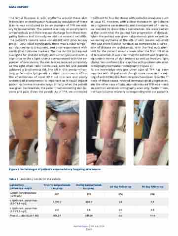
Figure 1. Serial images of patient’s extramedullary fungating skin lesions.
Table 1. Laboratory trends for the patient.
Laboratory (reference range)
Prior to talquetamab ramp-up
During talquetamab ramp-up
28 day follow-up
50 day follow-up
Lactate dehydrogenase (<240 u/L)
297
876
506
288
κ light chain, serum free (3.3-19.4 mg/L)
1,059.2
929.2
24
1.1
λ light chain, serum free (5.7-26.3 mg/L)
2.9
2.8
2.5
2.4
Free κ/ λ ratio (0.26-1.65)
365.24
331.86
9.6
0.46
Haematologica | 109 July 2024
2369
