
Page 41 - Haematologica July
P. 41
haematologica | 2018; 103(7)
Setd2 regulates hematopoietic stem cells
ly decreased Annexin V+ fractions and increased G0 frac-
tions in all 3 drug-treated groups compared with PBS treat-
ed group (Figure 7C-F), which indicates that SEC complex
inhibitors could partially rescue the HSC functional defi-
ciencies in Setd2Δ/Δ cells. However, the in vitro single-cell Setd2 loss leads to the significant upregulation of Nsds,
ABC
D
E
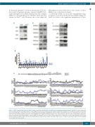
Figure 6. Setd2Δ/Δ hematopoietic stem cells (HSCs) show increased Nsds and RNA Pol II elongation associated phosphorylation changes. (A) Nsd1/2/3 and β-actin levels were determined by immunoblotting using bone marrow (BM) LSK cells. (B and C) H3K36me1/2, H3K4me3, H3K79me2, H3K27me3, H3, RNA pol II (Ser2P), pol II (Ser5P), Gata1, Gata3, Klf1, Myc, and β-actin levels were determined by immunoblotting using BM LSK cells from Setd2f/f and Setd2f/f/Vav1-Cre mice. (D) Relative gene expression levels were determined by qrt-PCR using flow sorted SLAM-HSCs from Setd2f/f and Setd2f/f/Vav1-Cre mice. Representative data were from 3 independent experiments. [N=6 each genotype; mean±Standard Error of Mean (SEM)]. (E) ChIP-qPCR assays of Setd2, Setd2 related histone modifications, and pol II phosphorylated forms [Pol II (Ser5P) and pol II (Ser2P)] on Myc locus was determined with c-kit+ BM cells from Setd2f/f and Setd2f/f/Vav1-Cre mice. (Setd2f/f=5 and Setd2f/f/Vav1-Cre=8; mean±SEM from 2 independent experiments).
differentiation assays failed due to the toxicity of those inhibitors in a long-term treatment.
Collectively, based on our results and published data, we propose a model for Setd2 function in HSCs (Figure 8).
1119
