
Page 34 - Haematologica July
P. 34
1112
Y. Zhou et al.
A
B
C
D
E
G
F
H
I
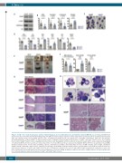
Figure 1. Setd2Δ/Δ mice showed leukopenia, anemia, erythroid dysplasia, increased thrombopoesis and mild bone marrow (BM) fibrosis. (A) Setd2 and H3K36me3 protein levels were determined by immunoblotting using c-Kit+ BM cells. Representative data were from 3 independent experiments. [N=3; mean±Standard Deviation (SD)]. (B) Complete blood count of Setd2f/f/Vav1-Cre mice, showing reduced white blood cells, lymphocytes, neutrophils, and platelets. [N=8 mice per genotype; mean±Standard Error of Mean (SEM)]. (C) Representative photos of Wright’s stained peripheral blood smear of Setd2f/f and Setd2f/f/Vav1-Cre mice. (D) Complete blood count of Setd2f/f/Vav1-Cre mice, showing reduced red blood cells, hemoglobin content, red blood cell specific volume (HCT), mean corpuscular hemoglobin concentration (MCHC), but increased mean corpuscular volume of red cells (MCV) and mean corpuscular hemoglobin (MCH). (N=8 mice per genotype; mean±SEM). (E) Representative photos of bones (tibia and fibula), spleens, and thymuses in Setd2f/f and Setd2f/f/Vav1-Cre mice. (F) BM cellularity, spleen weight, and thymus weight of Setd2f/f and Setd2f/f/Vav1-Cre mice. (N≥4 mice per genotype; mean±SEM). (G) Representative photos of hematoxylin & eosin-stained sections from the ster- num, spleens, thymuses of Setd2f/f and Setd2f/f/Vav1-Cre mice. (H) Dysplastic erythroid cells can be found in BM cytospin: megaloblastic erythroid precursors, dys- platic erythroid precursors with multi-nucleation, nuclear fragments, or nuclear budding. In addition, erythroid cells can be caught in mitosis. (I). Representative pho- tos of reticulin-stained sections from sternum of Setd2f/f and Setd2f/f/Vav1-Cre mice.
haematologica | 2018; 103(7)
