
Page 142 - Haematologica July
P. 142
1220
C. Lam et al.
cent imaging, beginning 13 days after implantation and on the same day as treatment initiation. Mice were treated for four weeks with vehicle or tofacitinib as indicated (five mice/arm.) Tofacitinib was formulated in 50% DMSO, 10% (polyethylene glycol 400) PEG 400, and 40% water and administered at 21.5 mg/kg daily by continuous subcutaneous infusion. All mouse studies were performed according to UCSF Institutional Animal Care and Use Committee-approved protocols.
Patient samples
De-identified primary MM BM samples were obtained from the UCSF Hematologic Malignancy Tissue Bank in accordance with the UCSF Committee on Human Research-approved proto- cols and the Declaration of Helsinki. BM mononuclear cells were isolated by density gradient centrifugation Histopaque-1077 (Sigma Aldrich), then adjusted to 2 x 10⁵/well in a 96 well plate. Primary cells were stimulated with 50 ng/ml recombinant human IL-6 (ProsPec) for 17 hours before treatment with tofacitinib for 24 hours. Cells were then stained with Alexa-Fluor 647 mouse anti- human CD138 antibody (BD Pharmingen) and SYTOX Green (Thermo) and analyzed on a CytoFLEX instrument (BD).
Results
Tofacitinib targets the BM microenvironment and reverses BMSC-mediated growth promotion
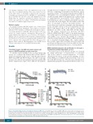
To initially validate findings from our drug repurposing screen, we cocultured the human MM cell line MM.1S, which was included in the screen,8 with the immortalized BMSC lines HS5 and HS27A (Figure 1A) and low-passage stromal cells derived from primary myeloma patient BM (Online Supplementary Figure S1C). MM.1S cell numbers
strongly increased compared to monoculture growth after 24 hours, confirming stromal-induced proliferative signal- ing in this cell line. Tofacitinib treatment reduced MM.1S cell numbers in a dose-dependent manner, such that at >1 mM tofacitinib, MM.1S cell numbers in coculture return to approximately monoculture levels (Figure 1A). Tofacitinib has no effect on MM.1S cell viability alone nor on stromal cells alone (Figure 1B). We further studied the effect of tofacitinib on several other MM cell lines. In monoculture we found that tofacitinib only demonstrates strong anti-MM activity in the IL-6 dependent cell line INA-6, with limited effect on the AMO-1 cell line and minimal to no effect on the other MM cell lines (Figure 1C). We further evaluated four myeloma cell lines (MM.1S, RPMI-8226, JJN-3, AMO-1) in which luciferase was stably expressed, allowing for the distinction of MM cell viability versus stromal cell viability in coculture.21 Only the stromal-responsive cell lines exhibit any sensitiv- ity to tofacitinib treatment (Figure 1D). Taken together, these results suggest that tofacitinib selectively targets the growth-promoting interaction between MM cells and the stromal microenvironment known to occur in patients.
BMSC-mediated plasma cell proliferation is through a mechanism partially dependent on IL-6
We next focused on the MM.1S cell line as it showed the unique phenotype of responsiveness to tofacitinib only in the context of stromal stimulation. To further characterize the nature of pro-growth signaling, we per- formed RNA-seq on MM.1S cells grown alone or in cocul- ture with HS5 stromal cells. We first noticed that the most significantly upregulated transcript in MM.1S in the cocul-
A
B
CD
Figure 1. Tofacitinib inhibits stromal cell-mediated proliferation in MM cells. A. Tofacitinib has no effect vs. MM.1S MM cells in monoculture, but instead reverses proliferation induced by BMSC lines HS5 and HS27A. B. Tofacitinib has no viability effect vs. BMSC C. Tofacitinib has limited or minimal effects vs. most MM cell lines in monoculture, except the IL-6 dependent line INA-6. D. In stromal cell coculture, tofacitinib does not have anti-MM effects vs. JJN-3 and RPMI-8226 cell lines, which do not proliferate in response to stroma, but shows strong reversal of proliferation in both MM.1S and AMO1 lines. All error bars represent +/- S.D. from CellTiter-Glo assay performed in quadruplicate in 384-well plates. PBS: phosphate buffered saline.
haematologica | 2018; 103(7)
