
Page 92 - Haematologica June
P. 92
S. Bertoli et al.
scriptomic characteristics in vivo. To test this, we used a patient-derived xenograft model of chemoresistance (Online Supplementary Figure S3A).19 Eight to 18 weeks after transplantation of the AML sample, the mice were given daily intraperitoneal injections of 60 mg/kg cytarabine or vehicle for 5 days. Three days after the last dose of cytara- bine or vehicle, viable human AML blasts were collected from the bone marrow, then purified and processed for transcriptomic analysis. The transcriptome of residual human AML cells exhibiting in vivo resistance to cytara- bine treatment displayed a strong upregulation of the genes involved in immune and inflammatory responses, including the nuclear factor-κB network (Figure 3E). Furthermore, this gene signature of chemoresistance dis- played a highly significant interaction with the dexam- ethasone gene signature (Figure 3F and Online Supplementary Table S5). Similarly, interrogation of a pub- licly available transcriptomic data set established from AML patients in first relapse and a data mining algorithm (Genomatix) revealed that the dexamethasone signature was also enriched within AML cells collected at relapse (Figure 3G and Online Supplementary Table S6).20 Moreover, in two patient-derived xenograft models, treatment of
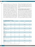
Table 3. Multivariate analysis for overall survival.
Numbers Events
NSG mice with the dexamethasone-cytarabine combina- tion induced a deeper therapeutic response compared to that achieved with cytarabine alone (Figure 3H). All together, these data strongly suggest that the impact of dexamethasone with intensive chemotherapy observed in the clinic could result from the targeting of chemoresistant AML cells.
Pre-clinical antileukemic activity of dexamethasone in acute myeloid leukemia with NPM1 mutations
A recent pre-clinical study demonstrated that AML cells with RUNX1 mutations were sensitive to glucocorticoids while earlier in vitro studies suggested an antileukemic activity in AML with the t(8;21)/RUNX1-RUNX1T1 translocation.21,22 To find out whether other molecular sub- groups could benefit from glucocorticoids, we first tested the in vitro activity of dexamethasone against AML cell lines with various genetic backgrounds. As expected, dex- amethasone had no significant activity as a single agent in most AML cell lines cultured in suspension (Figure 4A). Only two out of seven AML cell lines were moderately sensitive to the growth inhibition effect of dexametha- sone, including OCI-AML3, an NPM1-mutated cell line.
Dexamethasone
No 100 Yes 60
AML status
De novo 136 Secondary 24
Infection at diagnosis
No 124 Yes 33
Albumin- g/dL
>3.5 70 ≥3.5 88
Lactate dehydrogenase – UI/L
≤1550 80 >1550 80
Fibrinogen – g/L
≤1.5 15 >1.5 145
Cytogenetic risk
Favorable 15 Intermediate 127 Unfavorable 18
Hydroxyurea
No 52 Yes 108
Admission to intensive care unit*
No 114 Yes 46
Study period
2004-2009 67 2010-2015 93
Allogeneic stem cell transplantation
No 116 Yes 44
74 27
80
21
74 24
Adjusted HR
1 0.41
1
2.44
1 1.76
95% CI
0.22-0.79
1.45-4.11
1.06-2.93
0.39-0.94
1.14-2.70
0.17-0.62
1.28-13.62 1.83-27.29
0.39-0.97
2.21-6.38
0.36-1.07
P
0.007
0.001
0.029
0.027
0.010
0.001
0.018 0.005
0.037
<0.001
0.087
47 1
52 0.61
41
60 1.76
12 1
89 0.32 3 1
1
87 11
37
4.18 7.07
1
64 0.61
70
31 3.76
54
1
1
994
*During the first three months following chemotherapy. HR: Hazard Ratio; CI: Confidence Interval.
47 0.62
79
1
0.29-0.92
0.026
22 0.51
haematologica | 2018; 103(6)
