
Page 23 - Haematologica June
P. 23
Editorials
quito bites is an NK-cell proliferation that results from local and systemic symptoms following a mosquito bite. Immunophenotypic features, particularly CD30 expres- sion in a subset may overlap with features of lymphoma- toid papulosis.1,2,9,17 Recognition and timely diagnosis is important because patients with severe allergy to mos- quito bites may progress to systemic NK-cell type CAEBV, aggressive NK-cell leukemia, or ENKTL.17 In comparison to pediatric onset CAEBV, the adult cohort analyzed by Kawamoto et al. had significantly lower fre- quency of fever and greater frequency of skin lesions, including HV-LPD and severe allergy to mosquito bites.12
Distinguishing CAEBV from more aggressive EBV+ T- and NK-cell proliferations can be challenging, particularly given the propensity of CAEBV to progress to a more
aggressive LPD. It is, however, important to recognize and accurately diagnose LPDs in this continuum because early intervention may offer the only solution for patients progressing toward a fulminant disease. Therefore, defin- ing the boundary between CAEBV and more aggressive EBV+ T- and NK-LPDs, and the development of guidelines for the management of such patients, are urgently need- ed. In the Kawamoto et al. study, although no specific markers of disease severity or progression were identi- fied, thrombocytopenia (platelets <100x109/L), high EBNA titer (≥40), and HLH at initial diagnosis were asso- ciated with a worse overall clinical outcome. HLH was diagnosed using the HLH 2004 guidelines and was more frequent (46%) in the bone marrows of adult-onset CAEBV. Most importantly, allogeneic stem cell transplan-
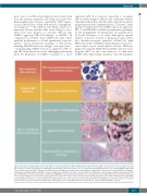
Figure1. Spectrum of Epstein-Barr virus-positive (EBV+) T-cell and natural killer (NK)-cell lymphoproliferative disorders (LPD). Characteristic features of EBV+ T-cell and NK-cell LPD are shown. Hemophagocytic lymphohistiocytosis (HLH) in the bone marrow of a 3-year old Asian child with EBV infection. Core biopsy highlights HLH within bone marrow sinuses (A and B). Chronic active EBV infection causing an oral cavity lesion in an otherwise asymptomatic 14-year old Caucasian adolescent showing a polymorphous lymphoid infiltrate with EBV+ cells in an angiocentric distribution (C and D). Systemic EBV T-cell lymphoma in an otherwise asymptomatic 8-year old child presenting with persistent fever and chills. A CD56 stain highlights the atypical infiltrate and underscores the importance of testing for EBV in a patient with unexplained fevers (E and F). Aggressive NK leukemia replacing the bone marrow in a 42-year old man who presented with pancytopenia and massive hepatosplenomegaly. EBER in situ hybridization shows an exuberant EBV+ interstitial infiltrate in the bone marrow (G and H). Extranodal NK/T-cell lymphoma, nasal type causing an ulcerated palatal lesion in a 51-year old man from Guatemala. Prominent necrosis and angiodestruction characterize the atypical lymphoid infiltrate (I and J).
haematologica | 2018; 103(6)
925
