
Page 178 - Haematologica June
P. 178
K. Kurakula et al.
SMCs (Figure 4C). These results indicate that Pin1 can sta- bilize TF, and that this stabilization is dependent on inter- action of Pin1 with the TFCD, but is not necessarily dependent on Pin1 isomerase activity.
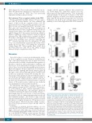
Pin1 enhances TF pro-coagulant activity via the TFCD Having established that Pin1 enhances TF gene expres- sion and TF protein half-life, we next examined the impact of Pin1 on the pro-coagulant activity of TF. We measured the Factor Xa-generating potential of the EC- RF24 cell line and primary human ECs and SMCs after Pin1 gain and loss-of-function. Pin1 overexpression markedly increased TF pro-coagulant activity in both ion- omycin-treated SMCs and TNF-α treated EC-RF24 cells and ECs (Figure 5A-C). In contrast, knockdown of Pin1 by siRNA or inhibition of Pin1 isomerase activity by Juglone attenuated the TNF-α induced pro-coagulant activity of TF in these cells (Figure 5A-C). Furthermore, using SMCs derived from wild-type mice and mice expressing the TFDCD truncated protein from the endogenous TF gene locus,23 we found that Pin1 can increase the activity of full- length TF, but not that of TF lacking the cytoplasmic domain (Figure 5D). Taken together, these results show that Pin1 interaction with TF via the TFCD strongly
increases the pro-coagulation activity of TF.
Discussion
Tissue Factor plays a crucial role in initiating the extrin- sic blood coagulation cascade. Therefore, detailed knowl- edge of the interacting proteins that modulate TF activity in vascular cells is essential to understand the regulation of thrombus formation under pathological conditions and may lead to novel intervention strategies for thrombotic disease. Here, we show that the peptidyl-prolyl isomerase Pin1 both enhances TF gene expression via activation of NF-κB and AP-1 signaling and directly interacts with TF through a well-conserved phosphorylated Ser258-Pro259 motif in its cytoplasmic domain. We elucidate the struc- tural details of this interaction and show that Pin1 increas- es both the protein half-life and pro-coagulant activity of TF in vascular cells. Additional effects of Pin1 on TF activ- ity may come from protease-activated receptor 2-induced release of TF on microvesicles, which was not studied in our experiments but were recently described.31
We demonstrate that Pin1 is a potent activator of TF gene expression in activated ECs and SMCs. The promot- er of the human TF gene contains transcription factor binding sites for NF-κB, AP-1, Sp-1 and Egr-1, which are essential for inducing TF expression in many cell types.9,24 Interestingly, Pin1 has been shown to bind and regulate the activity of all four of these transcription factors in a cell type-dependent manner.15 The deletion of Egr-1 and Sp1 response elements in the TF promoter constructs revealed that there is a complete abrogation of TF promoter activi- ty (data not shown). Therefore, the effect of Pin1 on activa- tion of the TF gene promoter by Egr-1 and Sp1 cannot be determined.
Concerning our detailed structural analyses, we con- clude that the TFCD-Pin1 WW-domain complex shows a larger contact area than what was observed in previous WW-domain/phosphopeptide NMR structures, but simi- lar to the RNAP II-CTD:Pin1 crystal structure.32 Residues in the anchoring zone of the Pin1 WW-domain-TFCD
complex showed apparent chemical shift perturbation (e.g. Tyr23) (Figure 3A), while these residues were stag- nant in previous Pin1 NMR titrations.32 This discrepancy could potentially be the result of titration of the short pThr-Pro fragment of Cdc25 or tau in these experiments, rather than the full protein domain that was used here. Furthermore, the β1-β2 loop1 conformer shows structural similarity to the isolated ligand-free Pin1 WW-domain X-
Figure 5. Pin1 enhances Tissue Factor (TF) pro-coagulant activity via the twen- ty-amino acid cytoplasmic domain (TFCD). (A-C) Factor Xa generation as a measure of TF activity in human human umbilical vein endothelial cells (HUVECs) (A), EC-RF24 cells (B), or smooth muscle cells (SMC) (C) after either overexpression or knockdown of Pin1 or treatment with the Pin1 inhibitor Juglone for 16 hours (hrs) followed by serum-starvation and treatment with ion- omycin for 3 hrs (SMCs) or TNF-α for 16 hrs [EC-RF24 and endothelial cells (ECs)]. (D) Factor Xa generation as a measure of TF activity in mouse SMCs derived from wild-type mice (WT) or mice expressing TF∆CD from the endogenous TF gene locus after overexpression of Pin1 and stimulation with ionomycin for 3 hrs. (E) Summary of Pin1 effects on TF: Pin1 enhances the protein half-life and pro-coagulant activity of TF through interaction with the conserved pSer258- Pro259 motif in the TFCD and enhances the activity of the transcription factors AP-1 and NF-κB to increase TF gene expression. Data are shown as mean±Standard Error of Mean. P-values were calculated using two-tailed Student’s t-test (A-C) or two-way ANOVA (D). *P<0.05, **P<0.01, ***P<0.001. AU: arbitrary units.
A
B
C
D
E
1080
haematologica | 2018; 103(6)
