
Page 174 - Haematologica June
P. 174
1076
K. Kurakula et al.
HEK293T cells with inhibitors of NF-κB or AP-1 signaling (Figure 1D). Similarly, mutating either the NF-κB or AP-1 response element sequences to become non-functional, as previously described,24 greatly reduced the ability of Pin1 to activate the TF promoter in both HEK293T cells and SMCs (Figure 1E and F). Finally, neither a Pin1 mutant with a disrupted WW-domain (Pin1:W34A) nor a Pin1 mutant lacking isomerase activity (Pin1:K63A) could acti- vate the TF promoter in HEK293T cells or SMCs (Figure 1G and H).
Taken together, these results show that Pin1 enhances TF gene expression and TF promoter activation via NF-κB
and AP-1, and that this activation of the TF promoter is dependent on both Pin1 isomerase activity and a function- al WW-domain.
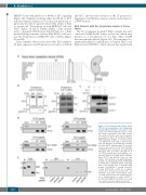
Pin1 interacts with the cytoplasmic domain of Tissue Factor
The TF cytoplasmic domain (TFCD) contains the well- conserved Ser258-Pro259 amino acid motif, which may function as a recognition site for Pin1 when Ser258 becomes phosphorylated (Figure 2A). We performed co- immunoprecipitation experiments in human SMCs and PMA-activated HUVECs, which showed that a pull-down
A
B
C
D
E
Figure 2. Pin1 interacts with Tissue Factor (TF) via the twenty-amino acid cytoplasmic domain (TFCD). (A) Amino acid sequence sim- ilarity of the TFCD in different species, show- ing strong conservation of the Pin1 recogni- tion motif Ser/Thr-Pro (box). (B) Co-immuno- precipitation (CoIP) for TF and Pin1 in human PMA-stimulated HUVECs or smooth muscle cells (SMC) with control IgG or anti-Pin1 anti- body (IP: IgG/Pin1) and analyzed by western blot with anti-TF antibody. Input TF and Pin1 levels are also shown. (C) CoIP for TF and Pin1 in HEK293T whole cell lysates over-expressing HA-tagged Pin1 and either full-length TF or TF∆CD using anti-HA antibody (IP: HA-Pin1) ana- lyzed by western blot with anti-TF antibody. Input TF/TF∆CD and HA-Pin1 levels are also shown. (D) Pull-down assay with biotinylated peptides encoding wild-type human TFCD (WT) or TFCD with an S258A mutation. Full-length Pin1, Pin1 mutants with a disrupted WW- domain (Pin1:W34A) or lacking isomerase activity (Pin1:K63A), or GFP (as a negative control) were detected by western blot. (E) Pull-down assay with cysteine-linked TFCD- encoding peptides that were unphosphorylat- ed or phosphorylated at either Ser253 or Ser258. Full-length Pin1 and GFP (as a nega- tive control) were detected by western blot.
haematologica | 2018; 103(6)
